1) Integumentary tissues
Integumentary tissues are located on the surface of plant organs. They consist of tightly closed cells and protect the internal parts of the plant from unfavorable external influences. Gives strength. According to their origin, they are distinguished from various meristems. primary And secondary covering tissues:
- Primary
Rhizoderm (epiblema) – primary single-layer superficial root tissue. The main function of the rhizoderm is absorption, selective absorption from the soil of water with mineral nutrition elements dissolved in it. Rhizoderm cells are thin-walled, with viscous cytoplasm and big amount mitochondria. Characteristic feature rhizoderm is the formation of some cells root hairs– tubular outgrowths . Root hairs increase the absorbing surface of the rhizoderm tenfold or more.
Epidermis- it covers leaves, stems of herbaceous and young shoots of woody plants, flowers, fruits and seeds. The main function of the epidermis is the regulation of gas exchange and transpiration(evaporation of water by living tissues). It prevents pathogens from entering the plant. The epidermis can function as an absorptive tissue. She takes part in the synthesis various substances, in the perception of stimuli, in the movement of leaves.
Composition of the epidermis:
Basic cells of the epidermis– living cells of tabular shape. The cells are tightly closed, intercellular spaces are absent. The side walls are often tortuous, which increases the strength of their adhesion; less often, they are straight.
Their inner, more powerful, layer consists of cellulose and pectin substances; the outer layer undergoes cutinization. A continuous layer of cutin is released on top of the outer walls, forming a protective film - cuticle.
Stomata– formations for the regulation of transpiration and gas exchange. Protects against dryness. The stomata consists of two trailing cells bean-shaped, between which there is stomatal gap, which can expand and contract. These cells often form hairs that develop into spines.
Lentils- formations in the form of small tubercles that serve for gas exchange.
- Secondary
Periderm–covers roots secondary structure and stems of perennial shoots. It can also occur as a result of healing of damaged tissue by the wound meristem.
The periderm consists of:
1)Cork consists of several layers of tabular cells, located densely, without intercellular spaces. The cork cells are dead, they do not have a protoplast and are filled with air. Substances (suberin) that increase the protective properties of the cork can also be deposited in the cell cavity. Main function plugs – protection against moisture loss.
2)Cork cambium– lays cork cells outside and cork parenchyma cells inside the organ. Cork parenchyma consists of living parenchyma cells. However, often the cork cambium works one-sidedly, depositing only the cork, while the cork parenchyma remains single-layered .
3) Crust. It occurs as a result of repeated formation of new layers of periderm in the deeper layers of the cortex. Thus, the crust consists of alternating layers of cork and other dead bark tissue. The thick crust reliably protects tree trunks from mechanical damage, etc.
Application: animal food.
2) Pollination- transfer of pollen from the stamens to the stigma of the pistil.
- Xenogamy- cross-pollination, in which the flowers of one plant are pollinated by pollen from the flowers of other plants of the same species.
Cross pollination device:
1) Dichogamy - non-simultaneous maturation in flowers anthers and stigmas.
2) Heterostyly - unequal length of columns of flowers on different plants the same type
- Cleistogamy- a type of self-pollination in which pollination occurs in closed flowers.
- Autogamy- self-pollination, in which pollen lands on the stigma of the same flower.
Features of self-pollination:
1) stamens and pistils mature at the same time
3) anthers above the pistil
3) anterior pollen is large, heavy
3) Angiospermsform one of the largest divisions of the plant kingdom. They make up the bulk of the plant mass in the biosphere. Angiosperms- Thisoak, birch, apple tree, wheat, rye, cabbage, palm tree, plantainetc. Many species of angiosperms are included in the number of cultivated plants.
Representatives of angiosperms grow everywhere. Some live very short - a few days. For example, the ephemerals spring krupka and Turchaninova breaker live 35-60 days and produce seeds. Others live hundreds of years. For example, the eastern plane tree, or plane tree, lives up to 2000 years, reaches 50 m in height, and its trunk is about 18 m in circumference.
In plants belonging to this division, the seeds are covered with the tissues of the fruit, which is formed from the ovary of the pistil of the flower. Thanks to these features, the department received the name Angiosperms or Flowering. Angiosperms (flowering) plants are extremely diverse in form and in requirements for living conditions, but all of them are characterized by general signs structure, reproduction and development.
The difference between two and one:
Dicotyledons: taproot, varied leaves, open wire tufts, five-membered flower. often reticulate veining. cotyledons (they contain food)
single: simple, linear, parallel veins, without stipules. vagina way attached, closed wire bundles, three-membered flower. in the endosperm there is food.
Integumentary tissues of the primary body of plants.
I. Epidermis. Leaves and young green shoots are covered, like a cover, with a single-layer primary integumentary tissue - the epidermis. Occasionally the epidermis is multilayered. Such multilayered epidermis was found in the leaves of the famous houseplant ficus (Ficus elastica). The epidermis arises from primary meristem - protodermis. It is a complex tissue because its cells vary in shape and, to some extent, in function. In particular, the cells that form stomata differ sharply from the cells of trichomes. The outer surface of the epidermal cells is often covered with a layer of cuticle or, less commonly, with a waxy coating of varying thickness. The cuticle can reach considerable thickness, especially in plants in arid habitats. Often its surface is covered various kinds folds or warty growths. With the exception of stomatal slits, the epidermal cells are tightly closed, i.e., there are no intercellular spaces. The main function of the epidermis is the regulation of gas exchange and transpiration, i.e. evaporation of water by the plant. Gas exchange and transpiration occur primarily through the stomata, but also partially through the cuticle. In addition, through the pores and strands of pectin substances in the outer walls cell membranes epidermis penetrates water and inorganic nutrients, which is especially typical for aquatic plants. Sometimes the epidermis performs functions unusual for this tissue - such as photosynthesis (in some aquatic plants), water storage (in some desert plants) or secretion of secondary metabolic substances (a number of essential oils).
The nature of the cells of the epidermis is different, the majority, called the main cells of the epidermis, are distinguished by a variety of outlines. The side walls, as a rule, are tortuous, which increases the density of their adhesion to each other, less often straight. The epidermal cells of the axial organs and leaves of many monocots are strongly elongated along the axis of the organ (Fig. 36).
In the main cells of the epidermis, a thin wall layer of protoplast with small sparse leucoplasts and a nucleus is found.
Often the entire cavity of the epidermal cell is occupied by one large vacuole. Its cell sap is colorless, but sometimes, especially in the epidermis of flowers and fruits, it is colored. The walls of epidermal cells are thickened unevenly. Usually the outer wall is thickest, while the side and inner walls are thin. Sometimes crystals are found in the cells of the epidermis; the cells of many cereals are impregnated with silica. The epidermal cells of many seeds contain polysaccharides in the form of mucus, which swells when moistened. The seeds easily stick to moving objects and thus spread.
Some plants have a special tissue under the epidermis - hypodermis. It partially performs a mechanical function,
partially protects the plant from excessive evaporation. A well-developed hypodermis is noticeable in the peculiar needle-like leaves - pine needles.
Stomata They are highly specialized formations of the epidermis, consisting of two guard cells, between which there is a kind of intercellular space, or stomatal fissure (Fig. 37). The gap can expand and contract by adjusting

transpiration and gas exchange. Under the gap there is a respiratory, or air, cavity, surrounded by cells of the leaf pulp. The epidermal cells adjacent to the trailing ones are called secondary, or parastomatal. They are involved in the movement of guard cells. Guard and side cells form stomatal apparatus. The number of side cells and their location relative to the stomatal fissure allows us to distinguish a number of stomatal types. They are being studied dentistry. Dentistry data are often used in plant taxonomy to clarify the systematic position of taxa. The most common stomatal types are shown in Figure 38.
Anomocytic the type of stomatal apparatus is common for all groups higher plants, excluding conifers. The side cells in this case do not differ from the rest of the epidermal cells. Diacitic the type is characterized by only two subsidiary cells, the common wall of which is perpendicular to the stomatal fissure. This type is found in some flowering plants, in particular in most Lamiaceae and Dianthus. At paracytic type, side cells are located parallel to the guard cells and the stomatal fissure. It is found in ferns, horsetails and a number of flowering plants. Anisocytic the type is found only in flowering plants. Here, the guard cells are surrounded by three side cells, one of which is noticeably larger or smaller than the others. Tetracite The type of stomatal apparatus is characterized mainly by monocots. At encyclocytic In this type, side cells form a narrow ring around guard cells. A similar structure is found in ferns, gymnosperms and a number of flowering plants. Location of guard cells
relative to other epidermal cells different types not the same. In some cases, guard cells are at the same level as the endidermal cells, sometimes they protrude above them or, on the contrary, lie much deeper (submerged stomata). The latter is observed in plants adapted to dry conditions. Sometimes the recesses in which the stomata are located are lined or covered with hairs. They are called stomatal crypts.
The number and distribution of stomata on a leaf or shoot vary depending on the plant species and living conditions. Their number usually ranges from several tens to several hundreds per 1 mm 2 of surface.
The mechanism of movement of guard cells is very complex and varies among different species. In most plants, when there is insufficient water supply at night, and sometimes during the day, the turgor in the guard cells decreases and the gap closes, thereby reducing the level of transpiration. With an increase in turgor, the stomata open. It is believed that the main role in these changes belongs to potassium ions. The presence of chloroplasts in guard cells is essential in the regulation of turgor. The primary starch of chloroplasts, turning into sugar, increases the concentration of cell sap. This promotes the influx of water from neighboring cells and the transition of guard cells to an elastic state.
The total area of stomatal openings is only 1-2% of the leaf area. Despite this, transpiration with open stomatal fissures reaches 50-70% of evaporation, equal in area to the open water surface.
Trichomes in plants, these are outgrowths of epidermal cells of different shape, structure and functions - hairs, scales, glands, nectaries. The sizes of trichomes vary widely. The longest trichomes (up to 5-6 cm) cover cotton seeds. Trichomes can be living or dead and serve a variety of functions. They are divided into covering and glandular. Coverts- These are unicellular, multicellular, branched and stellate hairs. Glandular trichomes(glands and nectaries) are elements of secretory tissues. The variety of covering trichomes is quite large (Fig. 39). Their structure and shape are sometimes used in taxonomy. Covering hairs form pubescence on plants of varying thickness, protecting against excessive transpiration or, occasionally, on the contrary, accelerating it. On leaves they most often appear on the side where there are stomata. The abundant pubescence of many desert plants helps to reflect the powerful solar radiation. Many tropical epiphytes use trichomes to absorb water and mineral salts.
In addition to hairs, on the epidermis of a number of species there are noticeable outgrowths called Emergents. These include the well-known stinging hairs of nettles, thorns of roses, raspberries, blackberries, etc. The thorns on the fruits of many umbellifers, datura, and chestnut are also emergent. Not only epidermal cells take part in the formation of emergers, but also the layers of cells lying underneath it.
II. Epiblema. Epiblema, often called rhizoderm, - primary single-layer integumentary tissue of the root. It arises from the outer cells of the apical meristem of this organ near the root cap and covers the young root endings. The epiblema is one of the most important plant tissues, since it is through it that water and mineral salts are absorbed from the soil.
In the root absorption zone, the epiblema is passive or active
absorbs elements of mineral nutrition, spending the latter case energy. In this regard, epiblema is rich in mitochondria. It is short-lived and, dying, transfers its functions to new areas of the epiblema of the growing root. The characteristics of epiblema cells correspond to the main function of the tissue. They are thin-walled, lack a cuticle and have a more viscous cytoplasm. It lacks stomata. Each epiblema cell is potentially capable of forming root hair, but more often root hairs are formed only from a part of the cells that have received a special name trichoblasts. Root hairs are usually unicellular, develop as a result of protrusion of the outer wall of the trichoblast and reach a length of 1-2 mm. They usually last for a few days and then die off.
The primary integumentary tissue of leaves and stems is the skin (epidermis). Knees -- living tissue, its cells contain a thin wall layer of protoplasm with a nucleus and plastids (most often with leukoplasts) and a large central vacuole.
The skin consists of tightly closed cells, usually having a sinuous outline in plan. In cross sections they are 4--5-angled, with the outer wall slightly convex. Skin cells of elongated organs (leaf petioles, leaf blades linear outline, stems), mostly elongated in a direction parallel to the longitudinal axis of the organ. In each skin cell, the outer wall is usually thickest. The side and inner walls are thinner and have pores. The shell consists mainly of cellulose. In many plants (especially grasses, sedges, and horsetails), the outer wall is encrusted with silica. The surface of the skin is covered with a film - cuticle (cuticle), consisting of cutan. On cross sections through the skin, cuticle spurs are often visible, penetrating into the side walls of the skin cells, and smaller spurs entering the cellulose thickness of the outer walls.
1 -- upper skin. 2 - cuticle. 3 - layer in the membranes of skin cells with cuticular plugs, 4 - cellulose layer, 5 - cell nucleus, 6 - pits in the skin (“external respiratory cavity”)
The leaf epidermis is the skin or covering tissue of the leaf. The epidermis consists of a single layer of flattened cells tightly adjacent to each other. In each cell, the main space inside is occupied by a central vacuole with cell sap. All organelles and the nucleus contained in the cell are pushed towards the membrane by the vacuole. However, the core, which is the custodian of all hereditary information, is clearly defined in each cell. The main cells of the leaf skin lack chloroplasts. Cells that have a different shape from the main ones and are arranged in pairs form stomata. Any stomata has a characteristic structure: it consists of two interlocking bean-shaped cells, and between these cells there remains a gap that resembles a lens in appearance. This intercellular space is called the stomatal fissure. The size and shape of the stomatal fissure are not constant. Its variability is determined by the closeness of the guard stomatal cells to each other. The structure of the guard stomatal cells differs from the structure of the main ones. The main difference is that stomatal cells contain chloroplasts in which photosynthesis occurs. The nucleus is also clearly visible in them, as in other cells of the leaf epidermis. The stomatal slits open to allow the air needed for photosynthesis and respiration to enter the leaf.
Stomata are highly specialized formations of the epidermis, consisting of two guard cells, between which there is a kind of intercellular space, or stomatal fissure. The gap can expand and contract, regulating transpiration and gas exchange. Under the gap there is a respiratory, or air, cavity, surrounded by cells of the leaf pulp. The epidermal cells adjacent to the trailing ones are called secondary, or parastomatal. They are involved in the movement of guard cells. Guard and side cells form the stomatal apparatus.
1 - guard cells,
2 - stomatal fissure,
3 - side cells,
4 - respiratory cavity,
5 - epidermal cells,
6 - cuticle
A - top view of the epidermis, B - cross-section of the stomatal apparatus
The total area of stomatal openings is only 1-2% of the leaf area. Despite this, transpiration with open stomatal fissures reaches 50-70% of evaporation, equal in area to the open water surface.
Epiblema (from the Greek epiblema - blanket, covering), rhizoderm, hair-bearing layer, primary superficial single-layer absorptive tissue of the root. Formed near the growth cone of the growing root from the dermatogen. Epiblema cells (trichoblasts) form outgrowths - root hairs, which greatly increase the absorbent surface of the root and anchor the plant in the soil, and also secrete a number of acids that act on the substrate substances. The epiblema is the first barrier tissue of the root, selectively absorbing soil substances. It is short-lived and, dying, transfers its functions to new parts of the epiblema of the growing root.
In the root absorption zone, the epiblema passively or actively absorbs elements of mineral nutrition, expending energy in the latter case. In this regard, epiblema is rich in mitochondria. It is short-lived and, dying, transfers its functions to new areas of the epiblema of the growing root.
The characteristics of epiblema cells correspond to the main function of the tissue. They are thin-walled, lack a cuticle and have a more viscous cytoplasm. It lacks stomata. Each epiblema cell is potentially capable of forming a root hair.
When growing freely - when roots develop in water or in moist air - the hairs have the shape of a cylinder or cone with a rounded end. Between solid particles In the soil, the hair becomes curved and in some places flattened or swollen in order to extract water and minerals from the soil.
Root hairs also play a mechanical role, providing support to the root tip, which breaks through the soil particles during growth, and facilitating anchoring
1.doc
^3.4. Integumentary tissues
Integumentary tissues are located on the surface of plant organs at the border with external environment. They consist of tightly closed cells and protect the internal parts of the plant from adverse external influences, excessive evaporation and drying, sudden changes in temperature, penetration of microorganisms, and serve for gas exchange and transpiration. According to their origin, they are distinguished from various meristems. primary And secondary covering tissues.
TO primary integumentary tissues include: 1) rhizoderm, or epibleme and 2) epidermis.
Rhizoderm (epiblema) – primary single-layer superficial root tissue. Formed from protodermis– the outer layer of cells of the apical meristem of the root. The main function of the rhizoderm is absorption, selective absorption from the soil of water with mineral nutrition elements dissolved in it. Through the rhizoderm, substances are released that act on the substrate and transform it. Rhizoderm cells are thin-walled, with viscous cytoplasm and a large number of mitochondria (mineral ions are actively absorbed, with energy consumption, against the concentration gradient). A characteristic feature of rhizoderm is the formation of some cells root hairs– tubular outgrowths, unlike trichomes, not separated by a wall from the mother cell ( rice. 3.4). Root hairs increase the absorptive surface of the rhizoderm tenfold or more. The hairs are 1-2 (3) mm long. Rhizoderm is often considered as suction textile.
Rice. 3.4. The tip of the root of Ozhika multiflorum: 1 – root hair.
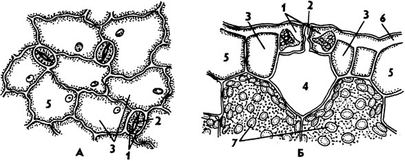
Epidermis- primary integumentary tissue formed from protodermis shoot growth cone. It covers leaves, stems of herbaceous and young shoots of woody plants, flowers, fruits and seeds. The main function of the epidermis is the regulation of gas exchange and transpiration(evaporation of water by living tissues). In addition, the epidermis performs a number of other functions. It prevents pathogens from entering the plant and protects inner fabrics from mechanical damage and gives organs strength. They can be released through the epidermis essential oils, water, salt. The epidermis can function as an absorptive tissue. It takes part in the synthesis of various substances, in the perception of irritations, and in the movement of leaves.
The epidermis is a complex tissue, its composition includes morphologically Various types cells: 1) main cells of the epidermis; 2) closing and subsidiary cells of stomata; 3) trichomes.
Basic cells of the epidermis– living cells of tabular shape. The appearance of cells from the surface is different ( rice. 3.5). The cells are tightly closed, intercellular spaces are absent. The side walls (perpendicular to the surface of the organ) are often tortuous, which increases the strength of their adhesion, less often straight. The epidermal cells of the axial organs and leaves of many monocots are strongly elongated along the axis of the organ. 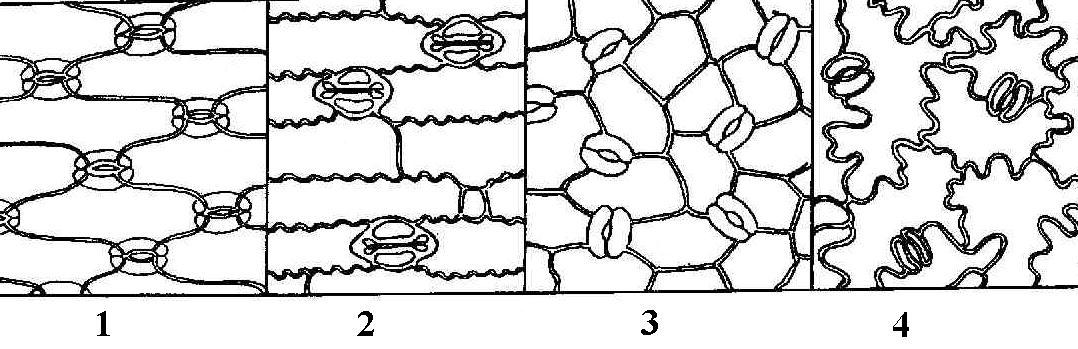
Rice. 3.5. Leaf epidermis various plants(view from the surface): 1 - iris; 2 - corn; 3 – watermelon; 4 - initial letter.
The outer cell walls are usually thicker than the rest. Their inner, more powerful, layer consists of cellulose and pectin substances; the outer layer undergoes cutinization. A continuous layer of cutin is released on top of the outer walls, forming a protective film - cuticle. In addition to cutin, it contains impregnations of wax, which further reduces the permeability of the cuticle to water and gases. Wax can be deposited in crystalline form and on the surface of the cuticle in the form of scales, rods, tubes and other structures visible only with an electron microscope. This bluish, easily erased coating is clearly visible on cabbage leaves, plums, and grapes. The power of the cuticle, the distribution of waxes and cutin in it determine the chemical resistance and permeability of the epidermis to gases and solutions. In dry climates, plants develop thicker cuticles. Plants immersed in water have no cuticle.
Epidermal cells have a living protoplast, usually with well-developed endoplasmic reticulum and the Golgi apparatus. Most plant species contain leucoplasts in the cytoplasm. Rare chloroplasts are found in aquatic plants, ferns, and inhabitants of shady places (hibiscus). The epidermis most often consists of a single layer of cells. Rarely, two- or multi-layered epidermis is found, mainly in tropical plants living in conditions of variable water supply (begonias, peperomia, ficus). The lower layers of the multilayered epidermis function as water-storing tissue. In some plants cell walls may be impregnated with silica (horsetails, cereals, sedges) or contain mucus (flax seeds, quince, plantain).
Stomata– formations for the regulation of transpiration and gas exchange. The stomata consists of two trailing cells bean-shaped, between which there is stomatal gap, which can expand and contract. Under the gap there is a large intercellular space - substomatal cavity. The epidermal cells adjacent to the guard cells are often different from the rest of the cells, and are then called side effects, or parastomatal cells(rice. 3.6). They are involved in the movement of guard cells.
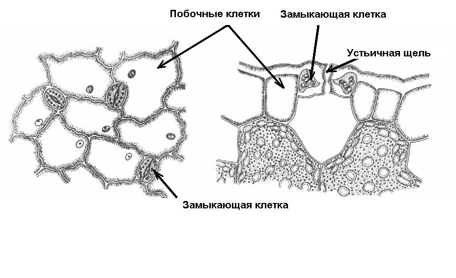
Rice. 3.6. Diagram of the structure of the stomata.
Guard and side cells form stomatal apparatus. Depending on the number of side cells and their location relative to the stomatal fissure, several types of stomatal apparatus are distinguished (Fig. 3.7). In pharmacognosy, types of stomatal apparatus are used to diagnose medicinal plant materials.
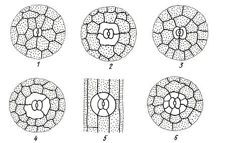
Rice. 3.7. Types of stomatal apparatus: 1 – anomocytic; 2 – diacite; 3 – paracytic; 4 – anisocytic; 5 – tetracite; 5 – encyclocytic.
Anomocytic the type of stomatal apparatus is common for all groups of plants, with the exception of horsetails. The side cells in this case do not differ from the rest of the epidermal cells. Diacitic the type is characterized by two subsidiary cells, which are located perpendicular to the stomatal fissure. This type is found in some flowering plants, in particular in most Lamiaceae (mint, sage, thyme, oregano) and cloves. At paracytic Typically, two side cells are located parallel to the guard cells and the stomatal fissure. It is found in ferns, horsetails and a number of flowering plants. Anisocytic the type is found only in flowering plants, in particular, it is found in cruciferous plants (shepherd's purse, yellowwort) and nightshade (henbane, datura, belladonna). In this case, the guard cells are surrounded by three side cells, one of which is noticeably larger or smaller than the others. Tetracite The type of stomatal apparatus is characterized mainly by monocots. At encyclocytic In this type, side cells form a narrow ring around guard cells. A similar structure is found in ferns, gymnosperms and some flowering plants.
The mechanism of movement of guard cells is based on the fact that their walls are thickened unevenly, so the shape of the cells changes as their volume changes. A change in the volume of cells of the stomatal apparatus occurs due to changes in osmotic pressure. The increase in pressure occurs due to the active intake of potassium ions from neighboring cells, as well as due to an increase in the concentration of sugars formed during photosynthesis. Due to the influx of water, the volume of the vacuole increases, turgor pressure increases, and the stomatal fissure opens. The outflow of ions occurs passively, water leaves the guard cells, their volume decreases, and the stomatal fissure closes. In most plants, stomata open during daylight hours and close at night. This is due to the fact that photosynthesis occurs only in light, and it requires an influx of carbon dioxide from the atmosphere.
The number and distribution of stomata vary greatly depending on the plant species and environmental conditions. In most plants their number is 100-700 per 1mm2 of leaf surface. With the help of stomata, the epidermis effectively regulates gas exchange and transpiration. If the stomata are completely open, then transpiration proceeds at the same rate as if there were no epidermis at all (according to Dalton’s law, for the same total area of the holes, the higher the evaporation rate larger number holes). When the stomata are closed, transpiration is sharply reduced and can actually only go through the cuticle.
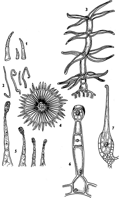
In many plants, the epidermis forms external single- or multicellular outgrowths various shapes – trichomes. Trichomes are extremely diverse, while remaining quite stable and typical for certain types, genera and even families. Therefore, the characteristics of trichomes are widely used in plant taxonomy and in pharmacognosy as diagnostic ones.
Trichomes are divided into: 1) coverts and 2) glandular. Ferrous Trichomes form substances that are considered secretions. They will be discussed in the section on excretory tissues.
^ Coverts trichomes look like simple, branched or stellate hairs, unicellular or multicellular ( rice. 3.8). Covering trichomes may long time remain alive, but more often they quickly die and fill with air.
A thick layer of hairs reflects part sun rays and reduces heating, creates a quiet space near the epidermis, which together reduces transpiration. Often the hairs form a cover only where the stomata are located, for example, on the underside of the leaves of coltsfoot and wild rosemary. Hard, prickly hairs protect plants from being eaten by animals, and papillae on the petals attract insects.
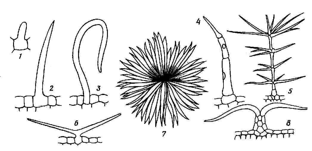
Rice. 3.8. Covering trichomes: 1-3 – simple unicellular, 4 – simple multicellular, 5 – branched multicellular, 6 – simple bicornuate, 7,8 – star-shaped (in plan and in cross-section of the leaf).
It is necessary to distinguish from trichomes, which are formed only from epidermal cells emergents, in the formation of which deeper tissues also take part. These include rose, raspberry, and blackberry thorns covering leaf petioles and young shoots.
TO secondary integumentary tissues include: 1) periderm and 2) crust, or rhytide.
Periderm- a complex multilayer integumentary tissue that replaces the primary integumentary tissues - rhizoderm and epidermis. The periderm covers the roots of the secondary structure and the stems of perennial shoots. It can also occur as a result of healing of damaged tissue by the wound meristem.
The periderm consists of three complexes of cells, different in structure and function. These are: 1) phellem, or cork, performing the main protective functions; 2) phellogen, or suberic cambium, due to the work of which the periderm as a whole is formed; 3) phelloderm, or cork parenchyma, which performs the function of feeding phellogen ( rice. 3.9).
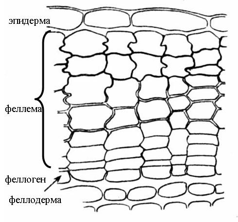
Rice. 3.9. The structure of the periderm of the elderberry stem .
Fellema (cork) consists of several layers of tabular cells, located densely, without intercellular spaces. Secondary cell walls are composed of alternating layers of suberin and wax, making them impermeable to water and gases. The cork cells are dead, they do not have a protoplast and are filled with air. Substances that increase the protective properties of the cork can also be deposited in the cell cavity.
^ Phellogen (cork cambium) – secondary lateral meristem. This is a single layer of meristematic cells that deposit plug cells on the outside and phelloderm cells on the inside of the organ. Phelloderm (cork parenchyma) refers to the main tissues and consists of living parenchyma cells. However, often the phellogen works one-sidedly, depositing only a plug, while the phelloderm remains single-layered ( rice. 3.9).
The main function of cork is to protect against moisture loss. In addition, cork protects the plant from the penetration of pathogenic organisms, and also provides mechanical protection to the trunks and branches of trees, and phellogen heals the damage caused, forming new layers of cork. Since the cork cells are filled with air, the cork case has low thermal conductivity and provides good protection against sudden temperature fluctuations.
In most trees and shrubs, phellogen is formed in annual shoots already in mid-summer. Most often it arises from parenchymal cells lying just below the epidermis ( rice. 3.9). Sometimes phellogen is formed in the deeper layers of the bark (currant, raspberry). Rarely, epidermal cells, dividing, turn into phellogen (willow, quince, oleander).
Gas exchange and transpiration in organs covered with periderm occur through lentils(rice. 3.10). In places where the lenticels are located, the cork layers are torn and alternate with parenchyma cells, loosely connected to each other. Gases circulate through the intercellular spaces of this performing tissue. Phellogen underlies the supporting tissue and, as it dies, is supplemented with new layers. With the onset of the cold season, phellogen is deposited under the performing tissue cap layer, consisting of cork cells. In spring, this layer breaks under the pressure of new cells. In the trailing layers there are small intercellular spaces, so that the living tissues of tree branches, even in winter, are not tightly separated from the environment.
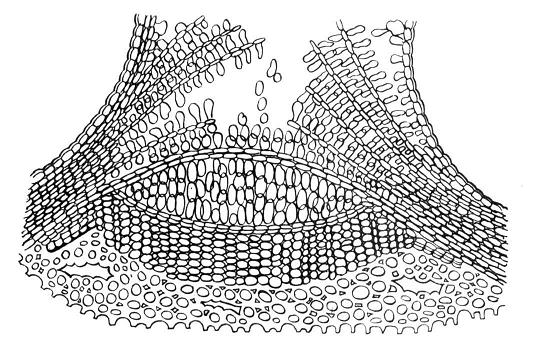
Rice. 3.10. The structure of the elderberry lentil in a cross section.
On young shoots, lentils look like small tubercles. As the branches thicken, their shape changes. In birch they stretch along the circumference of the trunk and form a characteristic pattern of black dashes on a white background. In aspen, the lenticels take the shape of diamonds.
In most woody plants, the smooth periderm is replaced by a fissured one. crust (rhytide). In pine this happens at 8-10 years, in oak - at 25-30 years, in hornbeam - at 50 years. Only some trees (aspen, beech, sycamore, eucalyptus) do not form a crust at all.
The crust arises as a result of the repeated formation of new layers of periderm in increasingly deeper layers of the cortex. Living cells enclosed between these layers die. Thus, the crust consists of alternating layers of cork and other dead bark tissue ( rice. 3.11).
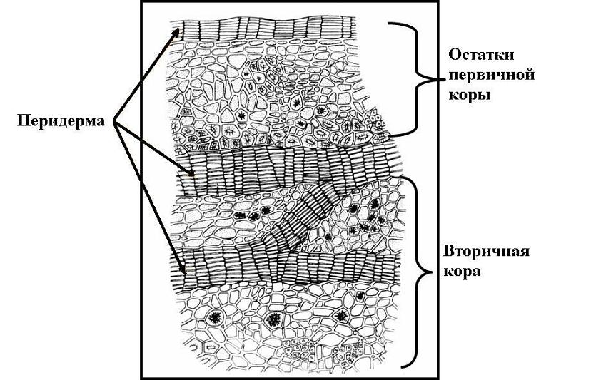
Rice. 3.11. Cross-section of oak bark .
Dead tissue the crusts cannot stretch, following the thickening of the trunk, so cracks appear on the trunk, but do not reach the deep living tissues. The boundary between the periderm and the bark is externally noticeable by the appearance of these cracks; this border is especially clear in birch, in which the white birch bark (periderm) is replaced by a black cracked bark. The thick crust reliably protects tree trunks from mechanical damage, forest fires, and sudden changes in temperature.