Integumentary tissues are located on the surface of plant organs at the border with external environment. They consist of tightly closed cells and protect the internal parts of the plant from unfavorable external influences, excessive evaporation and drying, sudden changes in temperature, penetration of microorganisms, serve for gas exchange and transpiration. According to their origin, they are distinguished from various meristems. primary And secondary covering tissues.
TO primary integumentary tissues include: 1) rhizoderm, or epibleme and 2) epidermis.
Rhizoderm (epiblema) – primary single-layer superficial root tissue. Formed from protodermis– the outer layer of cells of the apical meristem of the root. The main function of the rhizoderm is absorption, selective absorption from the soil of water with mineral nutrition elements dissolved in it. Through the rhizoderm, substances are released that act on the substrate and transform it. Rhizoderm cells are thin-walled, with viscous cytoplasm and big amount mitochondria (mineral ions are actively absorbed, with energy expenditure, against the concentration gradient). Characteristic feature rhizoderm is the formation of some cells root hairs– tubular outgrowths, unlike trichomes, not separated by a wall from the mother cell ( rice. 3.4). Root hairs increase the absorbing surface of the rhizoderm tenfold or more. The hairs are 1-2 (3) mm long. Rhizoderm is often considered as suction textile.
Rice. 3.4. The tip of the root of Ozhika multiflorum: 1 – root hair.
Epidermis- primary integumentary tissue formed from protodermis shoot growth cone. It covers leaves, stems of herbaceous and young shoots of woody plants, flowers, fruits and seeds. The main function of the epidermis is the regulation of gas exchange and transpiration(evaporation of water by living tissues). In addition, the epidermis performs a number of other functions. It prevents pathogens from entering the plant and protects inner fabrics from mechanical damage and gives organs strength. They can be released through the epidermis essential oils, water, salt. The epidermis can function as an absorptive tissue. She takes part in the synthesis various substances, in the perception of stimuli, in the movement of leaves.
The epidermis is a complex tissue, its composition includes morphologically Various types cells: 1) main cells of the epidermis; 2) guard and side cells of stomata; 3) trichomes.
Basic cells of the epidermis– living cells of tabular shape. The appearance of cells from the surface is different ( rice. 3.5). The cells are tightly closed, intercellular spaces are absent. The side walls (perpendicular to the surface of the organ) are often tortuous, which increases the strength of their adhesion, less often straight. The epidermal cells of the axial organs and leaves of many monocots are strongly elongated along the axis of the organ. 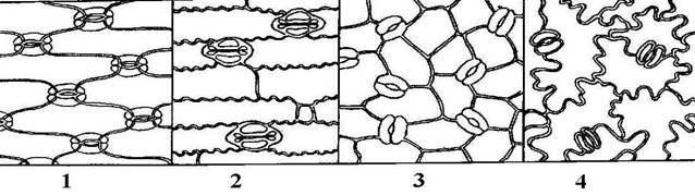
Rice. 3.5. Leaf epidermis various plants(view from the surface): 1 - iris; 2 - corn; 3 – watermelon; 4 - initial letter.
The outer cell walls are usually thicker than the rest. Their inner, more powerful, layer consists of cellulose and pectin substances; the outer layer undergoes cutinization. A continuous layer of cutin is released on top of the outer walls, forming a protective film - cuticle. In addition to cutin, it contains impregnations of wax, which further reduces the permeability of the cuticle to water and gases. Wax can be deposited in crystalline form and on the surface of the cuticle in the form of scales, rods, tubes and other structures visible only with an electron microscope. This bluish, easily erased coating is clearly visible on cabbage leaves, plums, and grapes. The power of the cuticle, the distribution of waxes and cutin in it determine the chemical resistance and permeability of the epidermis to gases and solutions. In dry climates, plants develop thicker cuticles. Plants immersed in water have no cuticle.
Epidermal cells have a living protoplast, usually with well-developed endoplasmic reticulum and the Golgi apparatus. Most plant species contain leucoplasts in the cytoplasm. U aquatic plants, ferns, inhabitants of shady places (hibiscus) there are rare chloroplasts. The epidermis most often consists of a single layer of cells. Rarely, two- or multi-layered epidermis is found, mainly in tropical plants living in conditions of variable water supply (begonias, peperomia, ficus). The lower layers of the multilayered epidermis function as water-storing tissue. In some plants cell walls may be impregnated with silica (horsetails, cereals, sedges) or contain mucus (flax seeds, quince, plantain).
Stomata– formations for the regulation of transpiration and gas exchange. The stomata consists of two guard cells bean-shaped, between which there is stomatal fissure, which can expand and contract. Under the gap there is a large intercellular space - substomatal cavity. The epidermal cells adjacent to the guard cells are often different from the rest of the cells, and are then called side effects, or parastomatal cells(rice. 3.6). They are involved in the movement of guard cells.

Rice. 3.6. Diagram of the structure of the stomata.
Guard and subsidiary cells form stomatal apparatus. Depending on the number of side cells and their location relative to the stomatal fissure, several types of stomatal apparatus are distinguished (Fig. 3.7). In pharmacognosy, types of stomatal apparatus are used to diagnose medicinal plant materials.
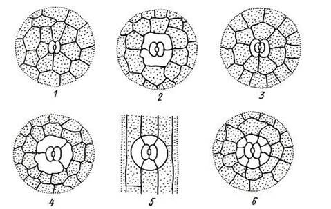
Rice. 3.7. Types of stomatal apparatus: 1 – anomocytic; 2 – diacite; 3 – paracytic; 4 – anisocytic; 5 – tetracite; 5 – encyclocytic.
Anomocytic the type of stomatal apparatus is common for all groups of plants, with the exception of horsetails. The side cells in this case do not differ from the rest of the epidermal cells. Diacitic the type is characterized by two subsidiary cells, which are located perpendicular to the stomatal fissure. This type is found in some flowering plants, in particular in most Lamiaceae (mint, sage, thyme, oregano) and cloves. At paracytic Typically, two side cells are located parallel to the guard cells and the stomatal fissure. It is found in ferns, horsetails and a number of flowering plants. Anisocytic the type is found only in flowering plants, in particular, it is found in cruciferous plants (shepherd's purse, yellowwort) and nightshade (henbane, datura, belladonna). In this case, the guard cells are surrounded by three side cells, one of which is noticeably larger or smaller than the others. Tetracite The type of stomatal apparatus is characterized mainly by monocots. At encyclocytic In this type, side cells form a narrow ring around guard cells. A similar structure is found in ferns, gymnosperms and some flowering plants.
The mechanism of movement of guard cells is based on the fact that their walls are thickened unevenly, so the shape of the cells changes as their volume changes. A change in the volume of cells of the stomatal apparatus occurs due to changes in osmotic pressure. The increase in pressure occurs due to the active intake of potassium ions from neighboring cells, as well as due to an increase in the concentration of sugars formed during photosynthesis. Due to the influx of water, the volume of the vacuole increases, turgor pressure increases, and the stomatal fissure opens. The outflow of ions occurs passively, water leaves the guard cells, their volume decreases, and the stomatal fissure closes. In most plants, stomata open during daylight hours and close at night. This is due to the fact that photosynthesis occurs only in light, and it requires an influx of carbon dioxide from the atmosphere.
The number and distribution of stomata vary greatly depending on the plant species and environmental conditions. In most plants their number is 100-700 per 1mm2 of leaf surface. With the help of stomata, the epidermis effectively regulates gas exchange and transpiration. If the stomata are completely open, then transpiration proceeds at the same rate as if there were no epidermis at all (according to Dalton’s law, for the same total area of the holes, the higher the evaporation rate larger number holes). When the stomata are closed, transpiration is sharply reduced and can actually only go through the cuticle.
In many plants, the epidermis forms external single- or multicellular outgrowths various shapes – trichomes. Trichomes are extremely diverse, while remaining quite stable and typical for certain types, genera and even families. Therefore, the characteristics of trichomes are widely used in plant taxonomy and in pharmacognosy as diagnostic ones.
Trichomes are divided into: 1) coverts and 2) glandular. Ferrous Trichomes form substances that are considered secretions. They will be discussed in the section on excretory tissues.
Coverts trichomes look like simple, branched or stellate hairs, unicellular or multicellular ( rice. 3.8). Covering trichomes may long time remain alive, but more often they quickly die and fill with air.
A thick layer of hairs reflects part sun rays and reduces heating, creates a quiet space near the epidermis, which together reduces transpiration. Often the hairs form a cover only where the stomata are located, for example, on the underside of the leaves of coltsfoot and wild rosemary. Hard, prickly hairs protect plants from being eaten by animals, and papillae on the petals attract insects.
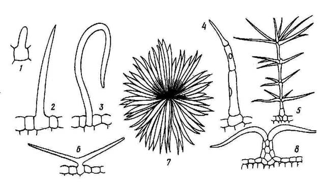
Rice. 3.8. Covering trichomes: 1-3 – simple unicellular, 4 – simple multicellular, 5 – branched multicellular, 6 – simple bicornuate, 7,8 – star-shaped (in plan and in cross-section of the leaf).
It is necessary to distinguish from trichomes, which are formed only from epidermal cells emergents, in the formation of which deeper tissues also take part. These include rose, raspberry, and blackberry thorns covering leaf petioles and young shoots.
TO secondary integumentary tissues include: 1) periderm and 2) crust, or rhytide.
Periderm- a complex multilayer integumentary tissue that replaces the primary integumentary tissues - rhizoderm and epidermis. Periderm covers the roots secondary structure and stems of perennial shoots. It can also occur as a result of healing of damaged tissue by the wound meristem.
The periderm consists of three complexes of cells, different in structure and function. These are: 1) phellem, or cork, performing the main protective functions; 2) phellogen, or cork cambium, due to the work of which the periderm as a whole is formed; 3) phelloderm, or cork parenchyma, which performs the function of feeding phellogen ( rice. 3.9).
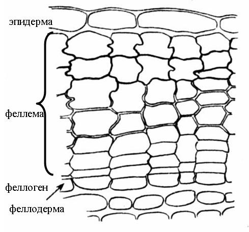
Rice. 3.9. The structure of the periderm of the elderberry stem .
Fellema (cork) consists of several layers of tabular cells, located densely, without intercellular spaces. Secondary cell walls are composed of alternating layers of suberin and wax, making them impermeable to water and gases. The cork cells are dead, they do not have a protoplast and are filled with air. Substances that increase the protective properties of the cork can also be deposited in the cell cavity.
Phellogen (cork cambium)– secondary lateral meristem. This is a single layer of meristematic cells that deposit plug cells on the outside and phelloderm cells on the inside of the organ. Phelloderm (cork parenchyma) refers to the main tissues and consists of living parenchyma cells. However, often the phellogen works one-sidedly, depositing only a plug, while the phelloderm remains single-layered ( rice. 3.9).
Main function plugs – protection against moisture loss. In addition, cork protects the plant from the penetration of pathogenic organisms, and also provides mechanical protection to the trunks and branches of trees, and phellogen heals the damage caused, forming new layers of cork. Since the cork cells are filled with air, the cork case has low thermal conductivity and provides good protection against sudden temperature fluctuations.
In most trees and shrubs, phellogen is formed in annual shoots already in mid-summer. Most often it arises from parenchymal cells lying just below the epidermis ( rice. 3.9). Sometimes phellogen is formed in the deeper layers of the bark (currant, raspberry). Rarely, epidermal cells, dividing, turn into phellogen (willow, quince, oleander).
Gas exchange and transpiration in organs covered with periderm occur through lentils(rice. 3.10). In places where the lenticels are located, the cork layers are torn and alternate with parenchyma cells, loosely connected to each other. Gases circulate through the intercellular spaces of this performing tissue. Phellogen underlies the supporting tissue and, as it dies, is supplemented with new layers. With the onset of the cold season, phellogen is deposited under the performing tissue cap layer, consisting of cork cells. In spring, this layer breaks under the pressure of new cells. In the trailing layers there are small intercellular spaces, so that the living tissues of tree branches, even in winter, are not tightly delimited from environment.
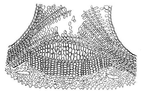
Rice. 3.10. The structure of the elderberry lentil in a cross section.
On young shoots, lentils look like small tubercles. As the branches thicken, their shape changes. In birch they stretch along the circumference of the trunk and form a characteristic pattern of black dashes on a white background. In aspen, the lenticels take the shape of diamonds.
In most woody plants, the smooth periderm is replaced by a fissured one. crust (rhytide). In pine this happens at 8-10 years, in oak - at 25-30 years, in hornbeam - at 50 years. Only some trees (aspen, beech, sycamore, eucalyptus) do not form a crust at all.
The crust arises as a result of the repeated formation of new layers of periderm in increasingly deeper layers of the cortex. Living cells enclosed between these layers die. Thus, the crust consists of alternating layers of cork and other dead bark tissue ( rice. 3.11).
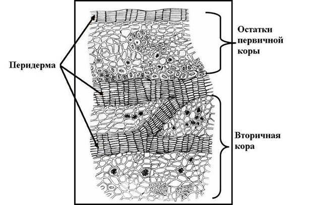
Rice. 3.11. Cross-section of oak bark .
Dead tissues of the crust cannot stretch, following the thickening of the trunk, so cracks appear on the trunk, but do not reach the deep living tissues. The boundary between the periderm and the bark is externally noticeable by the appearance of these cracks; this border is especially clear in birch, in which the white birch bark (periderm) is replaced by a black cracked bark. The thick crust reliably protects tree trunks from mechanical damage, forest fires, and sudden changes in temperature.
cover tissue plants are an outer tissue that protects plants from adverse environmental influences (temperature changes, drought, mechanical damage) and from various bacteria, viruses and fungi. Also, these tissues contribute to the absorption and release of water (which sometimes has to be retained), and carry out gas exchange.
Functions performed
Thus, the integumentary tissue performs the following functions:
- protective (separates internal environment plants from the external environment);
- exchange;
- excretory;
- receptor.
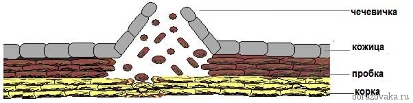
Figure 1. Plant integumentary tissues schematically
Kinds
There are such types of integumentary tissue as
- primary(located: epidermis - in the stem, exoderm - in the root);
- secondary or plug(also located in the roots and stems of plants - periderm; it usually appears after the epidermis dies);
- additional covering tissue or crust(rhythid).
The young root may also be covered with rhizoderm, similar in structure to the epidermis, but in general the epidermis and exoderm are dissimilar in structure and origin.
Epidermis- This living tissue, cork and crust- This dead tissue, the cells of which, dying, are filled with air and tannins, while continuing to perform their main functions - forming protective layers that protect the plant from adverse factors.
Structural features
All types of integumentary tissue are similar in structure (plant integumentary tissues are formed from cells and intercellular space) and have certain characteristics.
- The integumentary tissues contain a lot of cells and little intercellular substance.
- Cells and other structural particles are located very close to each other
- The integumentary tissue quickly regenerates (cells do not live long, they divide quickly, due to which the tissue is constantly renewed).
Structure and functions different types integumentary tissue is presented below in the table, which can be used in science lessons in 5th grade.
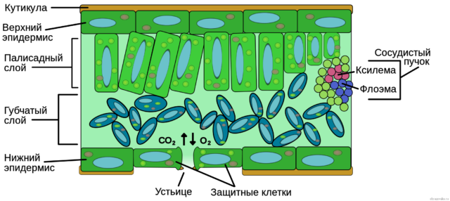
Fig 2. Plant epidermis
Types of plant integumentary tissue and their functions
| Type of covering tissue | Structure | Functions |
| Epidermis | U higher plants the entire surface of this tissue is covered with cuticle - a layer of cutin wax. There are special pores - stomata, which consist of several guard and side cells. Hairs or trichomes, consisting of one or more cells, are also present. |
This fabric provides gas and water exchange and regulates temperature. Hairs can perform protective functions(like, for example, nettle). |
| Periderm | It consists of three layers: phellem (outer layer or plug), phellogen (the main layer, the division of cells of which forms two other layers), phelloderm. This tissue contains so-called lentils, which promote gas exchange. | This tissue also protects the plant from pathogens. |
| Crust | Consists of dead phellogen cells. This fabric can stretch to certain limits, but it is not elastic, which is why cracks can gradually form on it. | This fabric is necessary to protect the plant from mechanical damage. Interestingly, the crust can even protect the plant from fire. This is why many trees survive forest fires. |
Fig 3. View of the cortex under a microscope
All fabrics can be either multi-layer or single-layer. For example, the rhizoderm covering the root is a single-layer tissue, while the epidermis is multi-layered. The periderm is also a complex, multi-layered integumentary tissue.
Some plant species, especially those that grow in arid areas, have another tissue (classified as primary) - velamen , which covers only the roots of the plant. It is this layer that ensures the timely supply of moisture to the leaves and stems of the plant.
Different tissues are formed in different period"life" of the plant. Thus, a crust can form in higher woody plants only in the 8th or 25th year of life (pine and oak, respectively). For some, it can even form in the 50th year of life (in hornbeam).
What have we learned?
Animals and plants have integumentary tissues. Plants have three types of integumentary tissue that perform different functions, although the main purpose of any covering tissue is to protect the plant. The structure of plant integumentary tissue is simple, but has its own characteristics. Fabrics can be single-layer or multi-layer. Some types of tissue begin to form when the plant reaches a certain age.