Plant cells are surrounded by a dense polysaccharide membrane, lined on the inside with plasmalemma.
Education cell wall occurs during metaphase and telophase of cell division. IN equatorial zone division, a middle plate appears, consisting of calcium pectate, which, growing from the center to the periphery, separates one newly formed cell from another. The middle plate is covered on both sides by the primary cell wall. Growth in thickness occurs due to the imposition of new layers from the contents of each cell. Cell growth in length begins with loosening of the matrix. In this process important role phytohormones play. New portions of material from which the cell wall is built enter the formed cavities. The synthesis and transport of these substances is carried out mainly by vacuoles of the Golgi apparatus.
The cell wall of cells dividing and growing by extension is called primary. After cell growth stops, new layers are deposited on the primary cell wall from the inside and a strong secondary cell wall.
The cell wall contains structural components(cellulose in plants, chitin in fungi), wall matrix components (hemicelluloses, pectin, proteins), encrusting components (lignin, suberin) and substances deposited on the wall surface (cutin and waxes). Cell walls may also contain calcium silicates and carbonates.
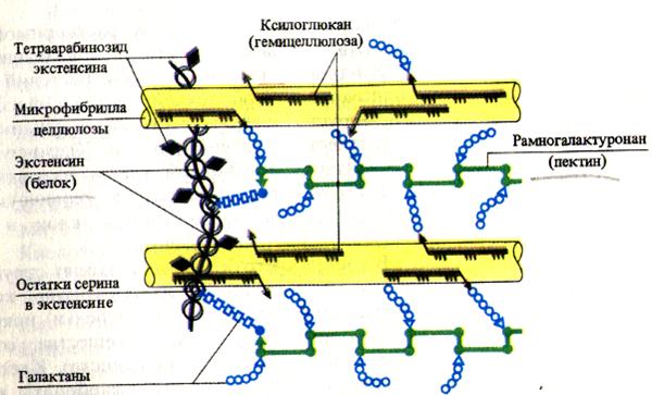
Cellulose(b-D-glucose polymer), hemicelluloses(polymers of hexoses and pentoses) and pectin substances(uronic acid derivatives) are carbohydrate components of cell walls. Cellulose and pectin substances adsorb water, providing hydration to the cell wall. Pectin substances containing many carboxyl groups bind divalent metal ions, which can be exchanged for other cations (H +, K +, etc.). This determines the cation exchange capacity of plant cell walls. In addition to carbohydrate components, the cell wall matrix also includes a structural protein called extensin. This is a glycoprotein containing more than 20% L-hydroxyproline of the total amino acids. According to this feature, the protein of plant cell walls is similar to the intercellular protein of animals - collagen.
Cellulose: A – structure of the cellulose molecule; B – cellulose molecule associations: 1 – micelle, 2 – microfibril, 3 – macrofibril
The main encrusting substance of the cell wall is lignin Intensive lignification of cell walls begins after cell growth ceases. Lignin is a polymer with an unbranched molecule consisting of aromatic alcohols (p-coumaric, coniferyl, synapic). The destruction and condensation of lignin in the soil is one of the factors in the formation of humus. Intensive lignification (impregnation of cellulose layers with lignin) of cell membranes begins after cell growth ceases. Lignin can be deposited in separate sections - in the form of rings, spirals or a network, as is observed in the cell walls of conducting tissue - xylem, or in a continuous layer, with the exception of those places where contacts are made between neighboring cells in the form of plasmodesmata. Lignin holds cellulose fibers together and acts as a very hard and rigid scaffold, enhancing the tensile and compressive strength of cell walls. It also provides cells with additional protection from physical and chemical influences, reduces water permeability. The lignin content in the shell reaches 30%. Encrustation of cell membranes with it leads to their lignification, which often entails the death of the living contents of the cell. Lignin combined with cellulose gives special properties wood, which make it an indispensable building material.
In the regulation of water and thermal regime plants involve tissues whose cell walls are impregnated suberin. The deposition of suberin makes the walls difficult to permeable to water and solutions (for example, in the endoderm, periderm). Suberin is deposited on the shell from the inside and makes it practically impermeable to water and solutions. As a result, the cell protoplast dies and the cell fills with air. This process is called suberization. Suberization of the cell membrane is observed in integumentary tissues perennial woody plants - periderm, cortex, as well as in the endodermis of the root. Suberin is the main substance of cork formations, to which these formations mainly owe their properties: impermeability to water, gases and low thermal conductivity.
The surface of plant epidermal cells is protected by hydrophobic substances - cutin And waxes: The precursors of these compounds are secreted from the cytoplasm to the surface, where their polymerization occurs. The cutin layer is usually penetrated by polysaccharide wall components (cellulose, pectin) and forms the cuticle. The cuticle is involved in the regulation of the water regime of tissues and protects cells from damage and infection.
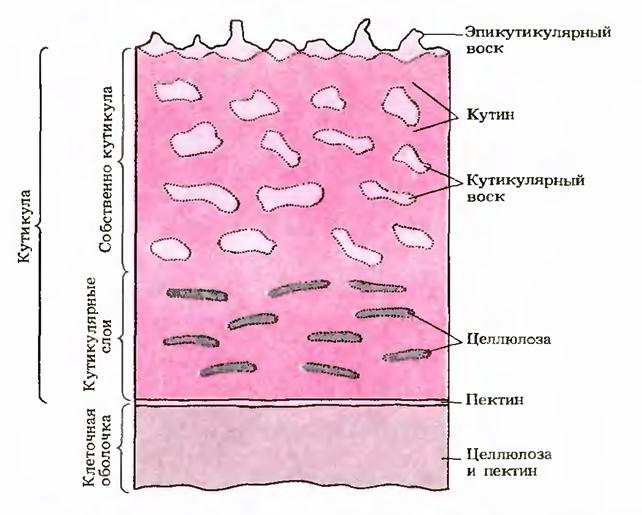
Accumulates in the membranes of the epidermal cells of some plants (cereals, sedges, etc.) a large number of minerals (mineralization), primarily calcium carbonate and silica. When mineralized, the leaves and stems of plants become tough, hard and less likely to be eaten by animals.
In primary cell walls, cellulose accounts for up to 30% of the dry mass of the wall. The amount of hemicelluloses and pectin substances varies depending on the object. Together with proteins, pectin substances can account for about 30% of the dry mass of the cell, with the amount of protein reaching 5 - 10%. About 40% comes from hemicelluloses.
1) Cell wall - structural education. Function: gives strength and shape, protects the protoplast from external conditions, participates in the conduction and absorption of substances.
The basis of the cell membrane (composition) is high-polymer carbohydrates (cellulose, i.e. fiber - is not digested, indicates low productivity), cellulose molecules are collected in complex bundles (mycelium), mycelium is combined into fibrils, their spaces are filled with hemicellulose (semi-fiber - less stable compound) and pectin (useful, swell in water, are a source of energy).
There are primary and secondary cell membranes. Meristematic and young growing cells have primary cell shell, thin, rich in pectin and hemicellulose; Cellulose fibrils in the matrix of the primary cell wall are arranged in a disorderly manner.
Secondary cellular The shell is usually formed when the cell reaches its final size and is superimposed in layers on the primary one from the protoplast side. In the secondary cell membrane, cellulose predominates; its fibrils are arranged in an orderly, parallel manner, but their direction in each layer is different, which increases the strength of the cell membrane. In the secondary cell wall there are openings (pores), where the cells are separated only by the primary cell wall and plasmodesmata (cytoplasmic bridges connecting neighboring plant cells).
Cell wall modifications:
- Lignification of the cell membrane occurs as a result of the deposition of lignin (a non-carbohydrate component in the fibrils); the cells lose their elasticity, but can allow water to pass through. These cells are more often dead than alive. Some cell walls may include: wax, cutina, suberin. Functions: gives the cell shape; separates one cell from another, is the skeleton for each cell and gives strength to the entire plant, performs a protective function.
- Cork formation is caused by a special fat-like substance - suberin. Such shells become impermeable to water and gases; they also do not allow heat to pass through; the contents of cells with suberized shells die.
- Cutinization involves the release of the fat-like substance cutin. Usually the outer walls of the skin of leaves and herbaceous stems are cutinized. This makes them less permeable to water, reduces the evaporation of water in plants, and protects them from overheating and ultraviolet radiation. Cutin forms a film on the surface of the organ called cuticle.
- Mineralization of cell membranes is the deposition of: silica and calcium salts. The cell membranes of the skin of leaves and stems of cereals, sedges, and horsetails are most heavily encrusted. The leaves of cereals and sedges can injure your hands.
- Mucusing of shells is the transformation of cellulose and pectin substances into mucus and gum. Mucilage is clearly observed on flax seeds that were in water. The formation of mucilage promotes better absorption of water by the seeds and their attachment to the soil.
2) Reproduction: the ability of a single individual to give rise to a whole series of its own kind.
Divided into: sexual and asexual (proper asexual and vegetative)
Vegetative: new individuals develop from individual vegetative organs or their interactions. It is carried out thanks to regeneration (the ability to restore an organism from a part of the body). Bio significance: the new organism is similar to the maternal one.
Methods of vegetative propagation:
- propagation by cuttings (a part of the plant that is not infected is planted in a substrate, sporodina),
- propagation by grafting (by sprouting parts of several plants, used in gardening),
- propagation by tubers (fleshy tubers with pita are planted in the ground, viviparous buckwheat),
- propagation by offspring (form shoots on roots, aspen),
- propagation by bulbs (in autumn they are planted from the plant itself into the ground)
- propagation by means of tendrils (creeping shoots, rooting, drupes, strawberries)
- propagation by rhizomes (underground shoot, pita stock, lily of the valley, violet, wheatgrass)
The use of vegetative propagation by humans. The rest is 40 cm.
WITH for a long time people, cultivating plants, began to use vegetative propagation. For example, growing potatoes, strawberries, banana in all countries of the world it is carried out only by vegetative means - tubers, tendrils and rhizomes.
The use of vegetative reproduction of plants in agricultural practice is called artificial vegetative propagation.
The main methods of artificial vegetative propagation come down to repeating those that occur in plants under natural conditions.
People often use propagation by cuttings - parts of green or woody shoots (grapes, currants, gooseberries, roses, cloves, ficus), tubers (potatoes, dahlia, sweet potato, Jerusalem artichoke), leaves (saintpaulia, gloxinia, begonia), bulbs (onion, garlic, tulip, daffodil), dividing the bush (currants, pyrethrum) and layering (gooseberries, honeysuckle, clematis), mustache (strawberry), rhizomes (sugar cane, irises, phlox), root shoots (plum, raspberry, cherry, lilac).
3) Pumpkin. Shape: herbs. Tap root. Stem: climbing, creeping, climbing Leaf: simple, petiolate, without stipules.
Formula: dioecious
1) regular female Ca (5) Co (5) A 0 G (3) perianth under the ovary
2) correct male Ca (5) Co (5) A 2+2+1 G 0
The inflorescence is solitary. Fruit: pumpkin
Representatives: cucumber, melon, pumpkin, watermelon, zucchini
Meaning: food, fodder
Cell membrane capable of thickening and modification. As a result of this, a veil is formed secondary structure. Thickening of the membrane occurs by applying new layers to the nerve membrane. Due to the fact that the imposition is already underway hard shell, cellulose fibrils in each layer lie parallel, and in adjacent layers - at an angle to each other. This achieves significant strength and hardness of the secondary shell. As the number of cellulose fibril layers increases and the wall thickness increases, it loses its elasticity and ability to grow. In the secondary cell wall, the cellulose content increases significantly, in some cases up to 60% or more. As cells continue to age, the membrane matrix may fill up various substances- lignin, suberin (woodiness or suberization of the shell). Lignin is formed from hemicellulose and pectin substances.[...]
The cell wall of wood fiber has several layers: primary, which is called the outer shell of the fiber, and secondary (the wall, which in turn consists of three layers: outer, middle and inner). Between the primary cell walls there is a layer of intercellular substance, through which the fibers are connected to each other. The secondary wall is relatively thick and represents the bulk of the cell volume.[...]
In the secondary layers of the cell walls of pine wood accumulated in large quantities mannans (22%) and uronic anhydride (25%).[...]
[ ...]
Cell wall thickening phase. How does thickening occur? During the growth period, the protoplast is surrounded only by the primary wall. When does a tree cell reach its largest size along the surface or shortly thereafter, the cell wall thickens. This is caused by the layering of the secondary wall on the primary, and this new layer arises as a result further activities protoplast inside the cell cavity. Naturally, cells in which the protoplast has disappeared cannot continue to thicken their walls. The formation of a secondary wall is a sign of an irreversible change in the cell, the further growth of which is already excluded, but further division is not necessarily excluded, provided that the daughter cells thus obtained occupy the same volume as the original cell.[...]
M.1ip - carpet, bedspread). It consists of tabular, thin-walled cells with dense cytoplasm. Usually it is single-rowed, but sometimes it is double-rowed or multi-rowed. Notoma cells are initially mononucleate, but later they often become binucleate or even multinucleate. The tapetum is a physiologically extremely active tissue: its cells contain enzymes, hormones, and nutritional material used in the process of microsporogesis. There are some reasons to consider the secretory type to be primary in evolutionary terms, and the amoeboid type to be secondary.[...]
It should be noted, however, that these data should be considered approximate, since the original preparations were not thoroughly purified.[...]
It is difficult to determine the cell wall location of polyuronide hemicelluloses because the reagents used to identify them also affect lignin. Some researchers suggest that hemicelluloses are the cementing substance between the fibrils and the various layers of the cell wall. Cohen even believes that secondary wall lignin has same nature with hemicelluloses. The basis for this assumption seems to be the fact that some carbohydrates, when processed strong acids may produce insoluble residues of a specific pattern. It should be emphasized, however, that areas, both carefully treated with reagents that dissolve hemicelluloses and not treated with them, give residues of a very similar structure when exposed to 72% sulfuric acid.[...]
To clarify the composition of individual layers of cell walls, an attempt was made quantification xylouronides in different layers of tracheids and libriform. Measurements were made on fibers from Japanese red pine, European fir, beech and birch. For this purpose, the fibers were carefully nitrated in a medium of acetic anhydride and carbon tetrachloride. Then the outer nitrated layer was removed by dissolving in acetone, after which the content of pentosans in the residue was controlled by furfural. It was found that pentosans in wood fibers are unevenly divided into layers. Largest quantity pentosans are found in the outer layers of fibers and their concentration decreases from the periphery to the center. Thus, the outer layers of coniferous wood fibers contain 50-80% pentosans, while hardwoods contain almost 100%. In the secondary layers of the cell walls of conifers, the content of pentosans turned out to be no more than 2-4%, and in deciduous trees it was 8-10%. Thus, chemical method confirmed the results obtained earlier by the ultraviolet light sorption method.[...]
A distinction is made between primary lignin, located in lignified cell walls (natural lignin) and secondary lignin - isolated lignin. The latter is largely a substance modified during the isolation process and contaminated with impurities of foreign substances. A change in lignin is expressed in the elimination of methoxyl groups, intramolecular condensation and other features.[...]
Many differences between tissue types are due to the structure of the cell wall, especially the secondary one. As we have already said, the formation of the primary cell wall occurs during the process of cell elongation, and, therefore, it must have the property of extensibility, while the secondary wall is formed after elongation has stopped.[...]
Preston
Simultaneously with these internal changes the outer hard wall of the oospore splits at its apex into five teeth, giving rise to a seedling emerging from the central cell (Fig. 269, 3). The first division of the central cell occurs by a transverse septum perpendicular to its long axis and leads to the formation of two functional various cells. From one, larger cell, a stem shoot is subsequently formed, which initial stage development is called a pre-adult, from another, smaller cell - the first rhizoid. Both of them grow by transverse cell divisions. The pre-adult grows upward and turns green quite quickly, filling with chloroplasts; the first rhizoid goes down and remains colorless (Fig. 269, 4). After a series of cell divisions, giving them the structure of single-row filaments, their differentiation into nodes and internodes occurs, and their further apical growth proceeds as described above for the stem. From the nodes of the pre-growth, secondary pre-shoots, whorls of leaves and lateral branches of the stem arise, from the nodes of the first rhizoid - secondary rhizoids and their whorled hairs. In this way, a thallus is formed, consisting of several stem shoots in the upper part and several complex rhizoids in the lower part (Fig. 2G9, 5).[...]
Supramolecular structure. Figure 6.10 shows a model of the structure of the cell wall. It includes 2 main layers: the primary wall P and the secondary. The latter is divided into 3 layers: 5], 5, Layer M, the middle plate, is an intercellular substance that connects cells to each other.[...]
In subsequent sections (part II) the chemistry of cell walls will be comprehensively discussed, relative quantities lignin in them and others related topics. However, concluding the consideration of the fourth and final phase of ontogenesis wood cell, it is worth mentioning some phenomena that are in one way or another connected with lignification, as botanists call it. Like the formation and proliferation of cells, as well as the thickening of cell walls, lignification can only occur during the life of the cellular protoplast, since dead cells cannot lignify their walls. The lignification process can be completed in the layer of intercellular substance and in the primary wall, but can continue in the secondary wall, even if this last-named layer is still centripetally increasing in thickness. In tree wood, lignification often ends very quickly in the layer adjacent to the inner side of the cambium, usually almost simultaneously with the time when the new cells have reached their largest size and the secondary walls have reached their final thickness. This explains why sapwood, at the same moisture content, is as strong or almost as strong as heartwood.[...]
Detailed study of the distribution of lignin and polysaccharides in the lignified cell walls of spruce and birch wood by measuring the absorption intensity of a thin beam ultraviolet rays when passing them through a transparent section, it confirmed the predominant location of lignin in the middle plate and primary wall, as well as partially in the outer layers of the secondary wall. In the middle plate of spruce wood, the lignin content reaches 73%, and in the secondary wall - no more than 16%. It follows that polysaccharides are concentrated mainly in the secondary layer. An attempt was made to measure by this method mutual arrangement cellulose and hemicelluloses. To do this, polysaccharides were first converted into colored compounds that absorb light.[...]
In most cells, alternating zones of greater or lesser lignin deposition are clearly visible, giving the appearance of concentric rings. In the opposite process, when the cell wall is treated with delignifying agents. reagents, the cellulose pattern remains the same. This indicates that there appear to be two interpenetrating systems, one consisting of cellulose and other polysaccharides, and the other of lignin. Bailey and Kerr showed that particle sizes reach 0.1¡x and less. The gaps or bands explain the relatively large "fibrils" seen by some researchers. In addition to the predominant concentric patterns, the grain of some types of wood exhibits an arrangement of radial lines or a combination of both types. Cells of compressed wood often have tough, almost solid bands of lignin near the cell cavity and radially arranged plates of it, separated by zones of polysaccharide substance, in the middle part of the cell wall.[...]
Lichens contain many elements and substances. All of them can be divided into two large groups- primary and secondary. Primary substances include those substances that directly participate in cellular metabolism; The body of lichens is built from them. Secondary products include the end products of metabolism, usually located on the walls of the hyphae. Many of these secondary lichen substances (in older literature they were called lichen acids) are specific to lichens and are not found in organisms from other systematic groups.[...]
Ritter, Lüdtke, et al. reported that when wood fibers are treated with various swelling agents, the secondary wall (and probably the primary wall as well) disintegrates into thread-like fragments or fibrils. Ritter divided these fibrils into spindle-shaped bodies, and these in turn into spherical units. The significance of such relatively large structural units(the length of the fusiform bodies is approximately 4[x) is unclear, due to the finely porous structure of the secondary wall described above. Neither the lignin residues after cellulose dissolution nor the cellulose residues after lignin dissolution show any noticeable gaps indicating the boundaries of these cell wall units. In addition, recent studies using an electron microscope have not established the presence of such relatively large units in the structure of cell walls.[...]
When assessing the effect of various wood-decaying fungi on plant tissue it is necessary to take into account that their individual hyphae. move selectively through cell walls. Thus, white rot fungi prefer the middle plate and primary shell, where lignin is mainly concentrated. Red or brown rot fungi, on the contrary, prefer to pass through the secondary shell, which is richest in carbohydrates. Accordingly, the color of the wood damaged by them also differs. These issues will be discussed in more detail later.[...]
Studies of tracheids and libriforms using a polarizing and electron microscope, as well as radiography, have established the existence of five concentric layers in the cell walls: the outer, or primary, wall and the secondary wall. The secondary wall in turn is divided into three layers, usually designated 81, vg and B3. In addition, between the primary walls of neighboring cells there is a middle plate that glues them together (Fig. 35).[...]
The increase in yields when using water vapor is explained by the fact that the removal of valuable products from the reaction space is accelerated and the development of secondary decomposition reactions is delayed. In addition, when water vapor comes into contact with the capillary system of wood on its surface layers, steam condensation is possible, which creates conditions for thermal decomposition in sour aquatic environment. In this case, decomposition reactions occur primarily in the layers of the cell wall, which are located with internal sides cell cavities and consist predominantly of non-heat-resistant hemicelluloses, which easily cleave off acetyl groups and part of the methoxyls associated with them, forming, respectively acetic acid and methyl alcohol.[...]
It is hardly correct to call the segments that make up the filaments of the spherople cells cells, not only because they have many nuclei and chloroplasts (and, therefore, are clearly secondary formations), but also because the transverse partitions separating them are not similar to the cell walls of other multicellular organisms. green algae. They vary greatly in shape, as well as in the method and place of formation (Fig. 226, 4-6). Often transverse septa take the form of ring-shaped internal thickenings on the cell walls that do not close in the center, so that a hole remains through which the cytoplasmic strand passes (Fig. 226, 4). In other cases, instead of partitions, special plugs are formed. And finally, groups of radially converging cords can appear anywhere in the thread, resembling the skeletal cords of the caulerpa and playing a mechanical role.[...]
Outside from plasma membrane their cells do not have an additional dense cell wall or it consists of chitin, rarely cellulose. Storage carbohydrates are usually in the form of glycogen (animal starch).[...]
Marx-Figipi and Pepcel studied the change in the DP of cotton pulp by various stages cotton ripening. They showed that the viscosity of cotton cellulose solutions decreased several hours after opening the box. Cellulose of the secondary cell wall in the fibers of unopened cotton bolls little maturity(cellulose yield - 18%) has a single maximum on the distribution curve at DP 14,000. About 10% of the material has a lower molecular weight (DP 1500-2500), this cellulose is contained in the primary cell wall.[...]
The position of the sites of microfibril formation in relation to the surface of the cytoplasmic membrane can be different. Thus, in bacteria this process occurs in an environment significantly removed from the cell surface and, therefore, from the membrane. Apparently, synthesis proceeds in a similar way in the thickened primary walls of the epidermal cells of oat coleoptiles, since cellulose synthesis in this case occurs evenly throughout the thickness of the cell wall. In the membranes of ascidians, cellulose deposition apparently also occurs in places remote from the surface of the secretory cells, although there is no sufficiently convincing evidence for this assumption. On the contrary, microfibrils of secondary plant cell walls are probably formed on the inner surface of the wall, in close proximity from the cytoplasmic membrane. Since there is much more cellulose in the secondary walls than in the primary walls, it can be concluded that the majority of cellulose microfibrils are formed near the cytoplasmic membrane. However, this is not mandatory.[...]
One of the methods based on this principle is the method of determining reactivity cellulose according to the pattern of swelling of xanthates in isopropyl alcohol. The swelling process during the interaction of fiber with solvent can be represented schematically in the following way: Liquid penetrates into the fiber, causing the volume of the fiber to increase. Then the weak elastic outer layer of the secondary cell wall of the fiber ruptures and swellings (“beads”) form at the sites of rupture. The remains of this layer form constrictions and cuffs on the swollen fiber. Then the outer layer is separated and the fiber swells evenly, transverse stripes are formed on it and the fiber is divided into packets of disks and separate disks, which subsequently dissolve.[...]
Dependence of wood strength on moisture content. Since the strength and stiffness of wood are determined in part by the cohesive forces that bind the molecules together, any agent that reduces these forces changes its overall strength. One such agent is water, so the strength of wood increases as the moisture content decreases, not only as a result of the increased density resulting from shrinkage, but also due to the presence of secondary valence cohesive forces1. Since the presence of water in an amount exceeding the saturation point of the fiber does not change the nature of the cell wall, the loss or acquisition of capillary (free) water has virtually no effect on the strength of wood. [...]
Structures containing a lot of lignin are dark brown to black, while weakly lignified areas are light yellow to amber. The results of this color reaction fully confirm previous work on cell wall chemistry. The secondary walls of the fibrous elements of hardwoods growing in temperate climates are lighter in color and therefore less lignified than the secondary walls of softwoods. The walls of blood vessels in deciduous trees are colored more dark color than the surrounding fibrous elements, therefore, they contain more lignin; the pore membranes are also highly lignified.[...]
This operation was carried out on lignified sections, previously freed from lignin using sodium chlorite in an acetic acid medium. The sections were then treated with p-phenylase; benzoyl chloride for the purpose of esterification of polysaccharides. Brightly colored orange-red color The sections were photometered after swelling in pyridine. By subjecting sections consisting of holocellulose to this treatment before and after removal of hemicelluloses, it was possible to establish that the bulk of hemicelluloses in spruce and birch wood are concentrated in the outer layers of the secondary wall. Thus, when extracting a cut of spruce holocellulose, 16% nsh caustic soda It was found that up to 60-80% of the alkali-soluble hemicelluloses of the total amount of polysaccharides are extracted from the outer layers of the cell, about 50% from the middle of the cell wall, and from the B3 layer. A similar picture was observed for cross sections of libriform from birch wood.[...]
Experiments by Ritter, and later by Bailey et al. showed that, regardless of the possible presence of pectic polyuronides in the middle plate, it consists mainly of lignin, as chemists understand it (insoluble in cold 72% sulfuric acid, soluble after chlorination and treatment weak grounds or basic salts). Moreover, Ritter proved that most of lignin is located in this layer. This statement contradicted the prevailing view at the time that most of the lignin was present in other layers, especially in the secondary wall. It was later proven that in such cases the seemingly wide and voluminous secondary wall is actually like a spider's web, which, after drying, shrinks and turns into scattered pieces. If the primary walls are included in a complex middle plate, then it is very likely that most of the lignin is located here. [...]
Calcium channels are also found in plant cell membranes. The regulation of the entry of 45Ca2+ microsomes isolated from corn coleoptils and pumpkin hypocotyls by light, PAA, and the dependence of this reaction on calmodulin was shown. For the functioning of voltage-gated Ca2+ channels (charophytic algae Lieu11op,m), the presence of Mg2+ is necessary. The state of these voltage-gated channels is controlled by a system of enzymes that monitor the level of cAMP in the cell. Data were also obtained indicating that direct action exogenous cAMP on the uptake of 45Ca2+ in the cells of the cyclone (mutant without a cell wall). The data shown in Fig. 4.1, indicate the regulatory effect of cAMP on the absorption of Ca2+ by cells. This indicates the possibility of mutual regulation of two systems of second messengers - cAMP and Ca2+. In experiments with animal cells, the increase in Ca2+ uptake under the influence of cAMP is explained by the phosphorylation of proteins of voltage-dependent Ca2+ channels and, as a result, an increase in their presence in the open state. [...]
Many studies have been devoted to studying the effect of ultrasound on cellulose fibers. Some researchers have compared or combined the effects of ultrasound with various mechanical influences. Thus, Yaime, Kronert and Neuhaus studied the effect of ultrasound on cellulose fibers in comparison with high-frequency mechanical vibrations and showed that ultrasound with a frequency of 20-3000 kHz loosens the fiber structure, increases the degree of its swelling and dehydration. The mechanical strength of paper made from such celluloses is increased, especially the tear strength. High-frequency ones act similarly. mechanical vibrations. Iwasaki, Lindberg and Meyer believe that the general pattern of changes in fiber structure under the influence of ultrasound in an aqueous environment is similar to changes in fiber structure during mechanical grinding. In this case, profound changes in the morphological structure of the fibers occur, leading to shifts in the secondary cell wall, separation of large pieces from the primary wall, then to swelling of the secondary wall and its defibrillation. In the work of Safonova and Klenkova, when studying microphotographs of fibers subjected to ultrasound in water, it was shown that there are other, deeper disturbances in the structure of the fiber, which becomes penetrated by a whole network of numerous transverse channels. It is noted that early wood fibers and fibers that have not been dried are more susceptible to ultrasound.
Plant cells, like the cells of prokaryotes and fungi, are enclosed in a relatively rigid cell wall. The material for the construction of this cell wall is secreted by the cell itself. living cell(protoplast). In terms of their chemical composition, plant cell walls differ from the cell walls of prokaryotes and fungi (Table 2.1), but these structures are characterized by some general functions, namely the functions of support and protection; in addition, both of them limit cell motility. The cell wall deposited during plant cell division is called the primary cell wall. Later, as a result of thickening, it can turn into a secondary cell wall. In this section we describe the process of formation of the primary cell wall. In Fig. 7.21 reproduces an electron micrograph in which you can see one of early stages this process.
Cell wall structure
The primary cell wall consists of cellulose microfibrils embedded in a matrix containing complex polysaccharides. Cellulose is also a polysaccharide (its chemical structure described in Sect. 5.2.3). Especially important for the role that cellulose plays in cell walls, it has a fibrous structure and high tensile strength comparable to that of steel. Individual cellulose molecules are long polysaccharide chains. Many such molecules cross-linked with each other hydrogen bonds, collected in strong bundles called microfibrils. Microfibrils immersed in the matrix form the framework of the cell wall. The cell wall matrix consists of polysaccharides, which for convenience of description are usually divided into pectins And hemicelluloses depending on their solubility in various solvents used for extraction. Pectins, or pectin substances, are usually isolated first during extraction because their solubility is higher. It is a mixed group of acidic polysaccharides (built from the monosaccharides arabinose and galactose, galacturonic acid, which belongs to the class of sugar acids, and methanol). Long molecules of pectin substances can be linear or branched. Median plate, which holds the walls of neighboring cells together, consists of sticky gelatinous pectates of magnesium and calcium. In the cell walls of some ripening fruits, insoluble pectin substances are converted back into soluble pectins. When sugar is added, these latter form gels; therefore they are used as gelling agents.
Hemicelluloses- this is a mixed group of polysaccharides soluble in alkalis (these include polymers of xylose, galactose, mannose, glucose and glucomanose). Hemicelluloses, like cellulose, have chain-shaped molecules, but their chains are shorter, less ordered, and more highly branched.
Cell walls are hydrated: 60-70% of their mass is usually water. Water moves freely through the free space of the cell wall. The presence of water affects chemical and physical properties cell wall polysaccharides.
Materials with increased mechanical strength, similar to the cell wall material, i.e., consisting of more than one component, are called composite materials or composites; their strength is usually higher than that of each of the components separately. Systems of fibers and a matrix (in engineering the basis of a composite material is called not a matrix, but a matrix. - Translation) are widely used in technology, so a lot of effort is spent on studying their properties both in technology and in biology. The compression matrix transfers stress to the tensile fibers. It also provides abrasive resistance and, apparently, resistance to adverse chemical influences that are possible in certain conditions. Reinforced concrete has long been used in construction, that is, a combination of concrete with steel reinforcement. Later, a lighter composite material appeared, in which the role of the matrix is played by plastic, and the role of reinforcement is played by glass or carbon fiber. Wood is a composite material; it owes its strength to its cell walls. An example of rigid composite materials biological origin Bone, cartilage, and the cuticle covering the exoskeleton of arthropods may also serve. There are also flexible composite materials, such as connective tissue.
Some cells, such as leaf mesophyll cells, have only a primary cell wall throughout their life. However, in most cells inner surface After the formation of the primary cell wall (outside the plasma membrane), additional layers of cellulose are deposited, i.e., a secondary cell wall appears. This usually occurs after the cell has reached its maximum size, and only a few cells, such as collenchyma cells, continue to grow during this phase. Secondary thickening of the plant cell walls should not be confused with secondary thickening (secondary growth) of the plant itself, i.e., an increase in the thickness of the trunk as a result of the addition of new cells.
In any layer of secondary thickening, cellulose fibers are located at the same angle, but in different layers this angle is different, which ensures even greater strength of the structure. This arrangement of cellulose fibers is shown in Fig. 7.27.
Some cells, such as tracheal xylem elements and sclerenchyma cells, undergo intense lignification(lignification); in this case, all layers of cellulose (primary and three secondary) are impregnated with lignin - a complex polymeric substance not related to polysaccharides. In protoxylem cells, lignin deposits have a ring, spiral or network shape, as can be seen in Fig. 8.11. In other cases, lignification is continuous, except for the so-called pore fields, i.e. those areas in the primary cell wall through which contact between neighboring cells occurs using a group of plasmodesmata (section 8.1.3 and Fig. 8.7). Lignin binds cellulose fibers together and holds them in place. It acts as a very hard and rigid matrix, enhancing the tensile and especially compressive strength of cell walls (prevents sagging). It also provides cells with additional protection from adverse physical and chemical influences. Together with cellulose remaining in the cell walls, lignin gives wood those special properties that make it an indispensable building material.
Functions of the cell wall
The main functions of plant cell walls are listed below.
1. Cell walls provide individual cells and the plant as a whole with mechanical strength and support. In some tissues, strength is enhanced by extensive lignification of the cell walls (small amounts of lignin are present in all cell walls).
2. The relative rigidity of cell walls and resistance to stretching determine the turgidity of cells when water enters them by osmosis. This enhances the supporting function in all plants and serves as the only source of support for herbaceous plants and for organs such as leaves, i.e. where secondary growth is absent. Cell walls also protect cells from rupture in a hypotonic environment.
3. The orientation of cellulose microfibrils limits and to a certain extent regulates both the growth and shape of cells, since the ability of cells to stretch depends on the location of these microfibrils. If, for example, microfibrils are located across the cell, surrounding it as if with hoops, then the cell into which water enters by osmosis will stretch in the longitudinal direction.
4. A system of cell walls interconnected with each other ( apoplast) serves as the main path along which water and minerals. Cell walls are attached to each other using median plates. The walls have small pores through which cytoplasmic strands pass, called plasmodesmata. Plasmodesmata bind the living contents of individual cells - they unite all protoplasts into unified system, in the so-called simplast.
5. The outer cell walls of epidermal cells are covered with a special cuticle film, consisting of a waxy substance called cutin, which reduces water loss and reduces the risk of pathogens entering the plant. In cork tissue, upon completion of secondary growth, the cell walls are impregnated with suberin, which performs a similar function.
6. The cell walls of xylem vessels, tracheids and sieve tubes (with sieve plates) are adapted for long-distance transport of substances throughout the plant. This issue is discussed in Chap. 8 and 14.
7. The walls of root endodermal cells are impregnated with suberin and therefore serve as a barrier to the movement of water (section 14.1.5).
8. In some cells, their modified walls store reserves nutrients; In this way, for example, hemicelluloses are stored in some seeds.
9. In transfer cells, the surface area of the cell walls is increased and the surface area of the plasma membrane is correspondingly increased, which increases the efficiency of transfer of substances by active transport(Section 14.8.6).
Cell wall formation begins during cell division. In the plane of division, a cell plate is formed, a single layer common to the two daughter cells. It consists of pectin substances having a semi-liquid consistency; there is no cellulose. In an adult cell, the cell plate is preserved, but undergoes changes, which is why it is called median, or intercellular plate (intercellular substance) (rice. 2.16). The median plate is usually very thin and almost indistinguishable.
Immediately after the formation of the cell plate, the protoplasts of the daughter cells begin to lay down their own cell wall. It is deposited from the inside both on the surface of the cell plate and on the surface of other cell walls that previously belonged to the mother cell. After division, the cell enters the elongation growth phase, which is caused by intense osmotic absorption of water by the cell associated with the formation and growth of the central vacuole. Turgor pressure begins to stretch the wall, but it does not tear due to the fact that new portions of microfibrils and matrix substances are constantly deposited into it. The deposition of new portions of material occurs evenly over the entire surface of the protoplast, so the thickness of the cell wall does not decrease.
The walls of dividing and growing cells are called primary. They contain a lot (60-90%) of water. The dry matter is dominated by matrix polysaccharides (60-70%), cellulose content does not exceed 30%, and there is no lignin. The thickness of the primary wall is very small (0.1-0.5 microns).
For many cells, cell wall deposition ceases simultaneously with the cessation of cell growth. Such cells are surrounded by a thin primary wall until the end of life ( rice. 2.16).
Rice. 2.16. Parenchyma cell with a primary wall.
In other cells, wall deposition continues even after the cell reaches its final size. In this case, the wall thickness increases, and the volume occupied by the cell cavity decreases. This process is called secondary thickening walls, and the wall itself is called secondary(rice. 2.17).
The secondary wall can be considered as additional, performing mainly a mechanical support function. It is the secondary wall that is responsible for the properties of wood, textile fiber, and paper. The secondary wall contains significantly less water than the primary wall; it is dominated by cellulose microfibrils (40-50% of the dry matter weight), which are located parallel to each other. Of the matrix polysaccharides, hemicelluloses (20-30%) are typical, and there are very few pectin substances. Secondary cell walls usually undergo lignification. In non-lignified secondary walls (flax bast fibers, cotton hairs), the cellulose content can reach 95%. Great content and strictly ordered orientation of microfibrils determine high mechanical properties secondary walls. Often, cells that have a secondary lignified cell wall die after the secondary thickening is complete.
The median lamina glues neighboring cells together. If it is dissolved, the cell walls lose contact with each other and separate. This process is called maceration. Natural maceration is quite common, in which the pectin substances of the middle plate are converted into a soluble state using the enzyme pectinase and then washed away with water (overripe fruits of pear, melon, peach, banana). Partial maceration is often observed, in which the middle plate does not dissolve over the entire surface, but only in the corners of the cells. Due to turgor pressure, neighboring cells in these places are rounded, resulting in the formation intercellular spaces(rice. 2.16). The intercellular spaces form a single branched network, which is filled with water vapor and gases. Thus, intercellular spaces improve gas exchange of cells.
A characteristic feature of the secondary wall is its uneven deposition on top of the primary wall, as a result of which unthickened areas remain in the secondary wall - pores. If the secondary wall does not reach a large thickness, the pores look like small depressions. In cells with a strong secondary wall, the pores in cross-section look like radial channels extending from the cell cavity to the primary wall. Based on the shape of the pore channel, there are two types of pores: simple and about edged(Fig. 2.17).
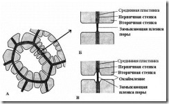
Rice. 2.17. Pore types: A – cells with secondary walls and numerous simple pores; B – a pair of simple pores; B – pair of bordered pores.
U simple pores The diameter of the pore channel is the same along its entire length and has the shape of a narrow cylinder. Simple pores are characteristic of parenchyma cells, bast and wood fibers.
Pores in two adjacent cells tend to appear opposite each other. These common pores look like one channel, separated by a thin partition of the middle plate and the primary wall. This combination of two pores of adjacent walls of neighboring cells is called pairs of pores and functions as one whole. The section of wall separating them is called closing film pores, or pore membrane. In living cells, the closing film of the pore is permeated with numerous plasmodesmata(rice. 2.18).
Plasmodesmata inherent only plant cells. They are strands of cytoplasm that cross the wall of adjacent cells. The number of plasmodesmata in one cell is very large - from several hundred to tens of thousands; usually plasmodesmata are collected in groups. The diameter of the plasmodesmal channel is 30-60 nm. Its walls are lined with plasmalemma, continuous with the plasmalemma of adjacent cells. In the center of the plasmodesmata there is a membrane cylinder - central rod plasmodesmata, continuous with element membranes endoplasmic reticulum both cells. Between the central rod and the plasma membrane in the canal there is hyaloplasm, continuous with the hyaloplasm of adjacent cells.
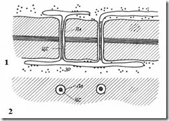
Rice. 2.18. Plasmodesmata under an electron microscope (diagram): 1 – on a longitudinal section; 2 – on a cross section; Pl– plasmalemma; CA– central rod of plasmodesmata; ER– element of the endoplasmic reticulum.
Thus, cell protoplasts are not completely isolated from each other, but communicate through plasmodesmata channels. They carry intercellular transport of ions and small molecules, and also transmit hormonal stimuli. Through plasmodesmata, protoplasts of cells in plant organism form a single whole called simplast, and the transport of substances through plasmodesmata is called symplastic Unlike apoplastic transport along cell walls and intercellular spaces.
U bordered pores(rice. 2.17) the channel sharply narrows during the deposition of the cell wall, so the internal opening of the pore, opening into the cell cavity, is much narrower than the external one, abutting the primary wall. Bordered pores are characteristic of early dying cells of water-conducting elements of wood. In them, the pore channel expands funnel-shaped towards the closing film, and the secondary wall hangs in the form of a roller over the expanded part of the channel, forming a pore chamber. The name bordered pore comes from the fact that when viewed from the surface, the internal opening looks like a small circle or narrow gap, while the outer hole, as it were, borders the inner one in the form of a circle of larger diameter or a wider slit.
Pores facilitate the transport of water and solutes from cell to cell without reducing the strength of the cell wall.