Ribosomes– ribonucleoprotein granules 25 nm in size. They consist of two subunits: small (10 nm) and large (15 nm), between which a strand of messenger RNA is located during protein biosynthesis (translation). In this case, the small subunit binds to RNA, and the large one catalyzes the formation of polypeptide chains. Ribosomal subunits are formed in the nucleoli and then exit the nucleus into the cytoplasm through nuclear pores. The assembly of ribosomes from their subunits occurs before the start of protein synthesis, and upon completion of the synthesis of the polypeptide chain, they disintegrate again.
A synthetically active cell contains several million ribosomes, which form about 5% of its dry mass. Distinguish free ribosomes (not associated with membranes and located in the hyaloplasm in suspension) and unfree ribosomes (associated with membranes of the cytoplasmic reticulum). Ribosomes can be located singly (in this case they are functionally inactive), but more often they are connected in chains that are strung, like beads, on thread-like messenger RNA molecules ( polyribosomes, polysomes). Free ribosomes synthesize proteins for the cell’s own needs, while non-free ribosomes synthesize proteins for export.
Cell center (cytocenter)– consists of two located perpendicular to each other centrioles.
Figure 2-8. Cell center (A) and centriole structure (B).
1. Centriole.
2. Satellites.
3. Triplet of microtubules.
4. Microtubules.
(According to V.L. Bykov).
Centriole is a hollow cylinder 200 nm thick and 300-500 nm long. The centriole wall is formed by 9 triplets of microtubules, 24 nm thick, built from globular protein tubulin. Neighboring triplets of microtubules are connected in the form of bridges by protein molecules dynein. Each triplet of microtubules is also associated with spherical structures - satellites. Microtubules diverge from the satellites to the sides, forming centosphere(Figure 2-8).
The cell center takes part in the formation of the division spindle; During mitosis, centrioles move toward the poles of the mother cell. In addition, centrioles take part in the formation of cilia and flagella.
Organelles of the cytoskeleton
The cytoskeleton is a complex dynamic three-dimensional network of microtubules, microfibrils and microfilaments, which provides: 1) maintenance and change in cell shape, 2) distribution and movement of cell components, 3) transport of substances into and out of the cell, 4) cell motility, 5) participates in in intercellular connections.
Microtubules have a thickness of 24 nm and a length of several microns. The microtubule wall thickness is 5 nm, and the lumen diameter is correspondingly 14 nm. They consist of 13 tubulin protofibrils running in a spiral. Microtubules are part of the spindle and ensure the divergence of chromosomes during mitosis, maintain the shape of the cell and ensure its mobility, and participate in the transport of macromolecules in the cell. Associated with microtubules is the protein kinesin, which is an enzyme that breaks down ATP and converts the energy of its breakdown into mechanical energy. At one end, the kinesin molecule is connected to a specific organelle, and at the other, with the help of ATP energy, it slides along the microtubule, moving the organelle in the cytoplasm.
Microtubules are a labile system in which the dissociation (destruction) of some microtubules and the assembly (formation) of others continuously occur. The site of microtubule formation ( microtubule organizing centers) are small spherical bodies satellites. They are located in the basal bodies of the cilia and the cell center.
Intermediate filaments (microfibrils) – protein filaments 8-11 nm thick. They form the framework of the cell, maintaining its shape and elasticity, and also ensure the orderly arrangement of organelles in the cell.
Microfilaments – protein filaments 5-7 nm thick. They are present in all cells and are located in its cortical layer (under the cytolemma). The composition of the proteins that form them in different cells different (actin, myosin, tropomyosin). They form the skeleton, frame of the cell, its intracellular contractile apparatus, ensure changes in the shape and movement of cells, cytoplasmic flow, endocytosis and exocytosis.
Functional systems (devices) of the cell- complexes of organelles that, under the control of the nucleus, ensure the performance of important cell functions. There are: 1) synthetic apparatus (includes endoplasmic reticulum, ribosomes, Golgi complex; 2 ) energy apparatus (mitochondria); 3) intracellular digestion apparatus vania(endosomes, lysosomes, peroxisomes); 4) cytoskeleton (microtubules, microfibrils, microfilaments, cell center).

 Organelles are permanent intracellular structures that have a specific structure and perform corresponding functions. Organelles are divided into two groups: membrane and non-membrane. Membrane organelles are presented in two variants: double-membrane and single-membrane.
 The double membrane components are plastids, mitochondria and cell nucleus. Single-membrane organelles include the organelles of the vacuolar system - the endoplasmic reticulum, Golgi complex, lysosomes, vacuoles of plant and fungal cells, etc. Non-membrane organelles include ribosomes and the cell center
 Single-membrane organelles Endoplasmic reticulum (ER) or reticulum - a complex system of channels and cavities various shapes(tubules, cisterns), penetrating the entire cytoplasm. a) Rough or granular endoplasmic reticulum: the membranes are covered with small granules - ribosomes. Functions: synthesis of polypeptides, their partial modification and transport b) Smooth, or agranular, endoplasmic reticulum: membranes are devoid of ribosomes, but enzymes of lipid and carbohydrate metabolism accumulate here. Functions: synthesis of lipids, steroids, carbohydrates, their transport.

 Functions: Connects all cell membrane structures in unified system. It is the surface on which all intracellular processes occur (synthesis of membrane proteins, lipids and carbohydrates). Spatially separates the cell. The transport of substances occurs through a system of channels.
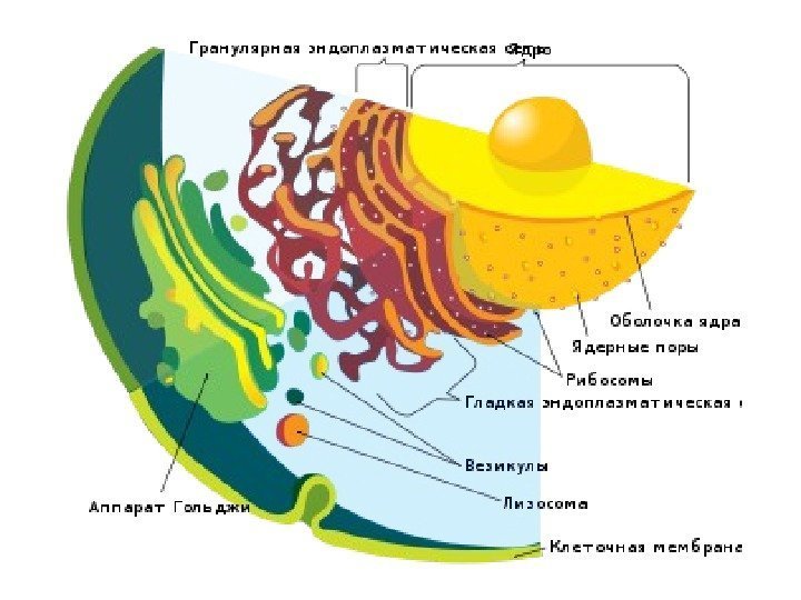
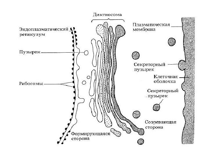
 The Golgi complex is present in almost all cells (with the exception of erythrocytes and sperm). Structure: A system of stacked flattened membrane sacs - cisterns, tubes and associated vesicles. Functions: Transport of substances, mainly proteins and lipids coming from the endoplasmic reticulum, their preliminary chemical rearrangement, accumulation, packaging into vesicles, formation of lysosomes.
 The Golgi apparatus was named after the Italian scientist Camillo Golgi, who first discovered it in 1897

 Functions of the Golgi Complex 1) sorting, accumulation and excretion of secretory products 2) completion of post-translational modification of proteins 3) accumulation of lipid molecules and formation of lipoproteins 4) formation of lysosomes 5) synthesis of polysaccharides for the formation of glycoproteins, waxes, mucus, matrix substances cell walls plants (hemicellulose, pectins) 6) formation of the cell plate after nuclear division in plant cells 7) participation in the formation of the acrosome; formation of contractile vacuoles of protozoa.
 Functions of the Golgi Apparatus: In the cisterns of the Golgi Apparatus, proteins intended for secretion, transmembrane proteins of the plasma membrane, lysosome proteins, etc. mature. The maturing proteins sequentially move through the organelle cisterns, in which their final folding occurs, as well as modifications - glycosylation and phosphorylation.
 Separation of proteins into 3 streams: 1. lysosomal - glycosylated proteins (with mannose) enter the cis-section of the Golgi complex, some of them are phosphorylated, and a marker of lysosomal enzymes is formed - mannose-6-phosphate. In the future, these phosphorylated proteins will not undergo modification, but will enter the lysosomes. 2. constitutive exocytosis (constitutive secretion). This flow includes proteins and lipids, which become components of the cell surface apparatus, including the glycocalyx, or they may be part of the extracellular matrix. 3. Inducible secretion - proteins that function outside the cell, the cell surface apparatus, during internal environment body. Characteristic of secretory cells.
![]() Concluding the consideration of the structure and operation of such a complex membrane organelle as the Golgi apparatus, it is necessary to emphasize that despite the apparent morphological homogeneity of its components, the vacuole and the cisterna, in fact, it is not just a collection of vesicles, but a slender, dynamic, complexly organized, polarized system.
Concluding the consideration of the structure and operation of such a complex membrane organelle as the Golgi apparatus, it is necessary to emphasize that despite the apparent morphological homogeneity of its components, the vacuole and the cisterna, in fact, it is not just a collection of vesicles, but a slender, dynamic, complexly organized, polarized system.

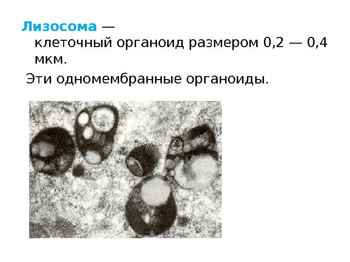
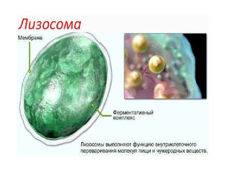
 Lysosomes Found in all cells, scattered throughout the cytoplasm. Structure: Single-membrane vesicles of various shapes and sizes; contain various proteolytic enzymes (about 40). Functions: Participate in intracellular digestion, i.e., the breakdown of large molecules. They can also destroy the structures of the cell itself, causing its death - autolysis.
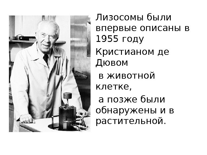 Lysosomes were first described in 1955 by Christian de Duve in animal cells, and were later discovered in plant cells.
 Lysosomes In plants, lysosomes are partly similar to lysosomes in their method of formation and in function. The presence of lysosomes is characteristic of cells of all eukaryotes. There are no uprokaryotic lysosomes, since they lack phagocytosis and do not have intracellular digestion.
 Signs of lysosomes One of the signs of lysosomes is the presence in them of a number of enzymes (acid hydrolases) capable of breaking down proteins, carbohydrates, lipids and nucleic acids.
 The enzymes of lysosomes include cathepsins (tissue proteases), acid ribonuclease, phospholipases, and others. Lysosomes contain enzymes that are capable of splitting off organic molecules sulfate (sulfatases) or phosphate (acid phosphatase) groups.
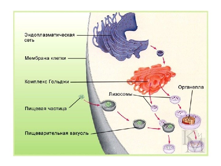
 Formation of lysosomes and their types Lysosomes are formed from vesicles (vesicles), separated from the Golgi apparatus, and vesicles (endosomes), into which substances enter during endocytosis. The membranes of the endoplasmic reticulum take part in the formation of autolysosomes (autophagosomes). All lysosome proteins are synthesized on sessile ribosomes. outside membranes of the endoplasmic reticulum and then pass through its cavity and through the Golgi apparatus.
 Lysosomes are heterogeneous organelles that have different shapes, sizes, ultrastructural and cytochemical features. "Typical" lysosomes in animal cells are usually spherical or oval shape. The number of lysosomes varies from one (a large vacuole in many plant and fungal cells) to several hundred or thousands (in animal cells).
![]() There are primary and secondary lysosomes. The former are formed in the region of the Golgi apparatus, they contain enzymes in an inactive state, while the latter contain active enzymes.
There are primary and secondary lysosomes. The former are formed in the region of the Golgi apparatus, they contain enzymes in an inactive state, while the latter contain active enzymes.
 Among lysosomes, one can also distinguish heterolysosomes (digesting material entering the cell from the outside through phago- or pinocytosis) and autolysosomes (destructing the cell’s own proteins or organelles).
 The most widely used classification of lysosomes and associated compartments is: Early endosome - it receives endocytic (pinocytotic) vesicles. Late endosome - vesicles with material absorbed during pinocytosis and vesicles from the Golgi apparatus with hydrolases enter it from the early endosome.
 classification Lysosome - vesicles with a mixture of hydrolases and digestible material enter it from the late endosome.
 Phagosome classification - it includes larger particles (bacteria, etc.) absorbed by phagocytosis. Phagosomes usually fuse with a lysosome.
 classification Autophagosome is a region of cytoplasm surrounded by two membranes, usually including some organelles and formed during macroautophagy. Fuses with the lysosome.
 classification Multivesicular bodies - usually surrounded by a single membrane, contain inside smaller vesicles surrounded by a single membrane. They are formed by a process reminiscent of microautophagy, but contain material obtained from the outside. The stage of formation corresponds to early endosomes.
 classification Residual bodies (telolysosomes) are vesicles containing undigested material (lipofuscin). In normal cells, they merge with the outer membrane and leave the cell by exocytosis. They accumulate with aging or pathology.
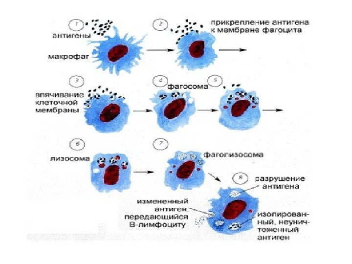
 Functions of lysosomes: digestion of substances or particles captured by the cell during endocytosis (bacteria, other cells); autophagy - destruction of structures unnecessary for the cell, for example, during the replacement of old organelles with new ones, or digestion of proteins and other substances produced within the cell itself
 autolysis is the self-digestion of a cell, leading to its death (sometimes this process is not pathological, but accompanies the development of the body or the differentiation of some specialized cells). Example: When a tadpole transforms into a frog, lysosomes located in the cells of the tail digest it: the tail disappears, and the substances formed during this process are absorbed and used by other cells of the body. Functions of lysosomes
 Clinical significance. Diseases associated with malfunction of lysosomes Sometimes, due to improper functioning of lysosomes, storage diseases develop, in which enzymes do not work or work poorly due to mutations. An example of storage diseases is amaurotic idiocy due to glycogen storage. The rupture of the lysosome and the release of hyaloplasma-splitting enzymes is accompanied by a sharp increase in their activity. This kind of increase in enzyme activity is observed, for example, in foci of necrosis during myocardial infarction and under the influence of radiation.
 Vacuole Vacuoles are single-membrane organelles that are “containers” filled with aqueous solutions organic and inorganic substances. The ER and Golgi complex take part in the formation of vacuoles. Young plant cells contain many small vacuoles, which then, as the cells grow and differentiate, fuse with each other and form one large central vacuole. The central vacuole can occupy up to 95% of the volume of a mature cell; the nucleus and organelles are pushed towards cell membrane. Membrane limiting plant vacuole, called tonoplast. The liquid that fills the plant vacuole is called cell sap. Part cell sap includes water-soluble organic and inorganic salts, monosaccharides, disaccharides, amino acids, final or toxic metabolic products (glycosides, alkaloids), some pigments (anthocyanins).
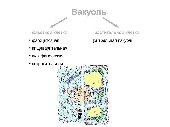 Vacuole animal cell plant cell phagocytotic digestive autophagic contractile Central vacuole
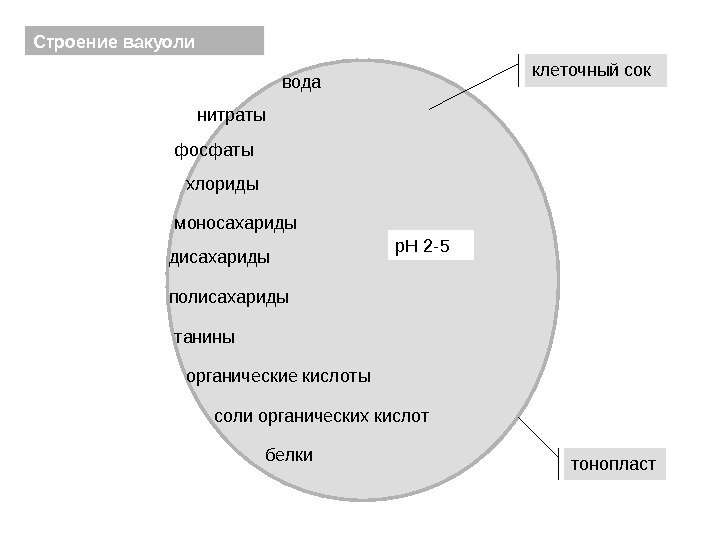 Structure of the vacuole tonoplast cell sap water nitrates phosphates chlorides monosaccharides disaccharides tannins organic acids salts organic acids p. H 2 -5 polysaccharides proteins
 Function No. 1 Maintaining turgor pressure. The vacuole functions as an osmometer and gives the cell the necessary strength and turgidity. Function No. 2 Sometimes vacuoles contain soluble pigments. This group includes anthocyanins, which are red, blue, or purple in color, and some related compounds that are yellow or cream in color. It is these pigments that mainly determine the color of flowers. Accumulation of reserve substances and “burial” of waste, i.e., the end products of cell metabolism. Sometimes vacuoles destroy substances that are toxic or unnecessary for the cell. Function No.
 Peroxisomes are tiny vesicles containing a set of enzymes. Functions: 1) Peroxisomes contain proteins on the surface of the membrane, which functions as a receptor recognizing signals on the introduced protein. Of all the peroxime proteins, the best known is the enzyme from the group of hydroperoxidases - catalase 2) Participate in metabolic reactions: in the metabolism of lipids, cholesterol, etc.
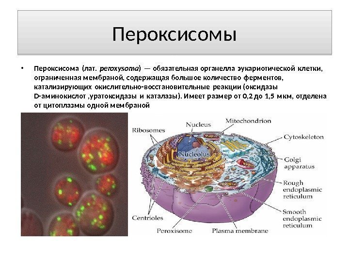 Peroxisomes Peroxisome (lat. peroxysoma) is an obligatory organelle of a eukaryotic cell, bounded by a membrane, containing a large number of enzymes that catalyze redox reactions (D-amino acid oxidases, urate oxidase and catalase). Has a size from 0.2 to 1.5 microns, separated from the cytoplasm by one membrane
 Functions of peroxisomes The set of functions of peroxisomes varies in cells different types. Among them: oxidation of fatty acids, photorespiration, destruction of toxic compounds, synthesis of bile acids, cholesterol, and ester-containing lipids, construction of the myelin sheath nerve fibers, phytanic acid metabolism, etc. Along with mitochondria, peroxisomes are the main consumers of O 2 in the cell. The peroxisome usually contains enzymes that use molecular oxygen to abstract hydrogen atoms from certain organic substrates to form hydrogen peroxide: Catalase uses the resulting one to oxidize a variety of substrates - for example, phenols, formic acid, formaldehyde and ethanol: This type oxidative reactions is especially important in liver and kidney cells, whose peroxisomes neutralize many toxic substances entering the bloodstream. Almost half of the ethanol entering the human body is oxidized to acetaldehyde in this way. In addition, the reaction has implications for the detoxification of the cell from hydrogen peroxide itself.
 Non-membrane organelles Ribosomes are found in all types of cells (including prokaryotic ones). They can lie freely in the cytoplasm or connect to the membranes of the ER. Found in mitochondria and plastids. Structure: Small spherical bodies formed by two unequal subunits - large and small, which consist of 3-4 molecules of ribosomal RNA and more than 50 protein molecules. Ribosomes always contain magnesium ions that support their structure. Functions: synthesis of polypeptide chains (the second stage of protein synthesis is translation).
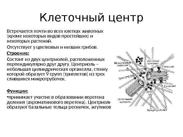 Cellular center Found in almost all animal cells (except some types of protozoa) and some plants. Absent in flowering and lower fungi. Structure: Consists of two centrioles located perpendicular to each other. The centriole is a small cylindrical organelle, the wall of which is formed by 9 groups (triplets) of three fused microtubules. Functions: takes part in the formation of the fission spindle (achromatin spindle). Centrioles form the basal bodies of cilia and flagella.
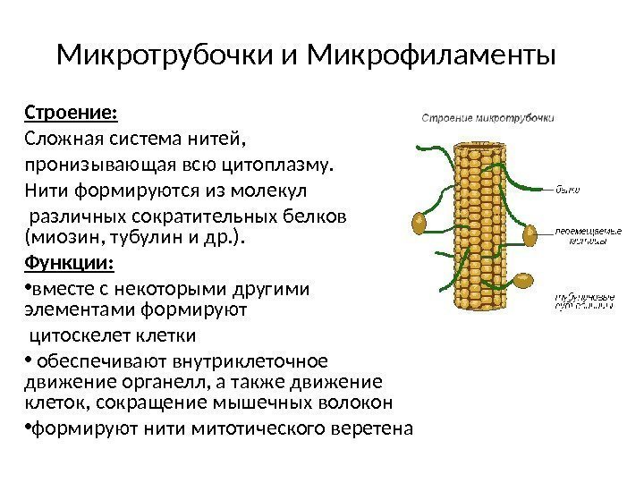 Microtubules and Microfilaments Structure: A complex system filaments that penetrate the entire cytoplasm. The threads are formed from molecules of various contractile proteins (myosin, tubulin, etc.). Functions: together with some other elements, they form the cytoskeleton of the cells, ensure intracellular movement of organelles, as well as cell movement, contraction of muscle fibers form the threads of the mitotic spindle
 Red - nucleus Green - microtubules Yellow - Golgi apparatus

Atlas: human anatomy and physiology. Complete practical guide Elena Yurievna Zigalova
Membrane organelles. Transport across membranes
Human cells are characterized by the presence of a huge number of intracellular membranes, forming several compartments (from the English compartment - “compartment, compartment”), differing from each other in structure and function: cytosol, nucleus, endoplasmic reticulum, Golgi complex, mitochondria, lysosomes, peroxisomes. Due to the presence of these elements, a large number of different biochemical reactions occur simultaneously in the cell.
All membrane organelles are built from elementary membranes, the structural principle of which is similar to the structure of the plasmalemma described above. The absorption of macromolecules and particles by cells occurs by endocytosis (from the Greek endon - “inside”, kytos - “cell”), release - by exocytosis (from the Greek exo - “outside”, kytos - “cell”).
One of essential functions The plasmalemma is transport. Let us recall that the hydrophobic “tails” of lipids facing each other prevent the penetration of polar water-soluble molecules. There are two types of transport: passive and active. The first does not require energy, the second is energy dependent. As a rule, the inner (cytoplasmic) surface of the membrane carries negative charge, which facilitates the penetration of positively charged ions into the cell. Water enters the cell by osmosis(from the Greek osmos - “push, pressure”), which is the slow penetration of water through a semi-permeable membrane separating two solutions of different concentrations. As a result, the concentrations of these two solutions are equalized.
Diffusion(from Latin diffusion - “spreading, spreading”) is the transition of ions or molecules caused by their Brownian motion through membranes from a zone where these substances are in a higher concentration to a zone with a lower concentration until the concentration is both sides of the membrane are aligned. Specific transport proteins built into the membrane transport small polar molecules across it, with each protein transporting one class of molecules or only one compound. Some transmembrane proteins form channels. Active transport carried out by carrier proteins, while energy is consumed resulting from the hydrolysis of ATP (adenosine triphosphate acid) or proton potential. Active transport occurs against a concentration gradient. To carry out biochemical reactions, substances must enter the cell through endocytosis and excretion of exocytosis metabolic products.
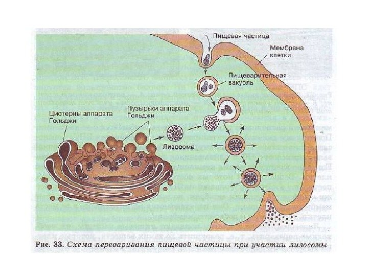
Endocytosis. There are several methods of endocytosis. The entry of liquid colloidal particles is called pinocytosis, and large particulate matter– phagocytosis. In order for external molecules to enter the cell, they must first be bound by glycocalyx receptors. The cytolemma begins to invaginate, then its edges come closer and close, splitting off the vesicle containing the trapped molecules. An endreome is formed, which is immersed in the cytoplasm and meets with lysosomes. Their membranes fuse. In the resulting secondary lysosome, substances entering the cell undergo breakdown.
Exocytosis ensures the removal of large molecular compounds. First, they segregate in the Golgi complex in the form of transport vesicles and are directed to the cell surface. The membrane of the vesicle is embedded in the cytolemma, and the contents of the vesicle appear outside the cell.
Two varieties are known endocytosis: phagocytosis – absorption of particles (from the Greek phagos - “devouring” and kytos - “cell”) and pinocytosis – absorption of dissolved substances (from the Greek Pino - “drink”). The phagocytosed particle enclosed in a membrane is called a phagosome. During the process of endo- and exocytosis, transported substances are enclosed in membrane vesicles.
Endoplasmic reticulum, or endoplasmic reticulum(ER), is a single continuous cavity bounded by a membrane that forms many invaginations and folds ( see fig. 1). Therefore, on electron diffraction patterns, the endoplasmic reticulum appears in the form of many tubes, flat or round cisterns, and membrane vesicles. There are two types of ER: granular and agranular. The side facing the cytosol of the first is covered with ribosomes, while the side of the second is devoid of them. Function of granular ER: synthesis of proteins by ribosomes and transport of proteins, smooth synthesis and exchange of carbohydrates and lipids (steroid hormones, glycogen, cholesterol) and neutralization (hepatocytes), synthesis of chlorides, from which it is formed in the stomach hydrochloric acid. Being a depot of calcium ions, smooth ER is involved in muscle contraction; delimits future platelets in megakaryocytes. One of the most important functions of the ER is the synthesis of membrane proteins and lipids for all cellular organelles.
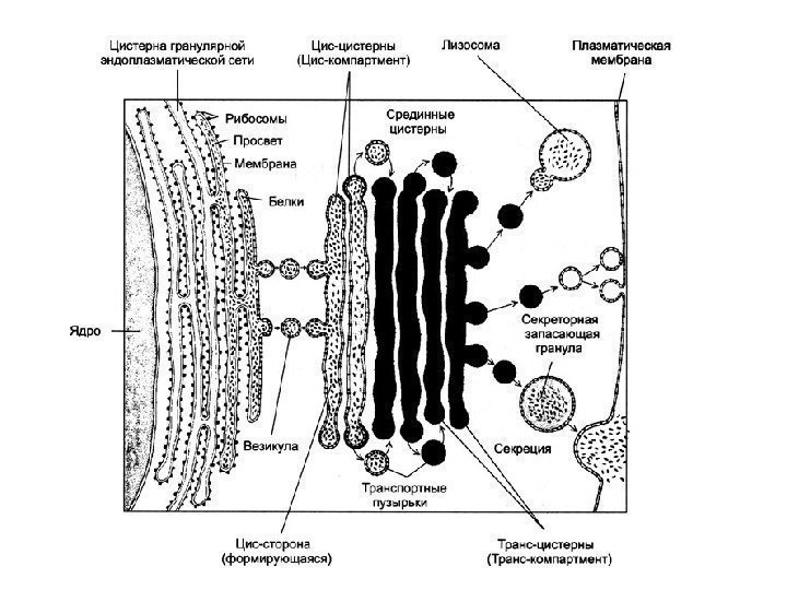
Complex or Golgi apparatus(CG), is a set of tanks, bubbles, plates, tubes, sacs, bounded by a membrane, in which synthesized products are accumulated and packaged ( see fig. 1). These products are removed from the cell with the help of the complex elements; in addition, polysaccharides are synthesized, protein-carbohydrate complexes are formed, and transported molecules are modified. In a light microscope, the CG appears in the form of a mesh or a system of tubules and vacuoles. CG is present in all human cells, except erythrocytes and horny scales of the epidermis. In most cells, the CG is located around or near the nucleus. In the CG, three membrane elements are identified: flattened sacs (cisterns), vesicles and vacuoles. CG is a three-dimensional cup-shaped structure consisting of several (from one to several hundred) dictyosomes (from the Greek dyktion - “network”). Each dictyosome contains 4–8 (on average 6) parallel flattened cisternae, penetrated by pores with widened ends, from which vacuoles containing synthesized substances are split off. The cisternae are associated with many membranous vesicles as well as larger secretory granules. Elements of the Golgi complex are interconnected by channels.
The membranes of the Golgi complex are formed and maintained by granular endoplasmic reticulum, in which membrane components are synthesized. They are transported by transport vesicles budding from the ER and merge with the CG, from which secretory vesicles are constantly budding, and the cisternae membranes are constantly renewed. They supply the glycocalyx and synthesized substances to the plasmalemma, thus ensuring the renewal of the plasmalemma. One of the most important functions of the CG is protein sorting.
Lysosomes- membrane organelles containing about 50 types of various hydrolytic enzymes, which are synthesized on the ribosomes of the granular endoplasmic reticulum, from where they are transferred by transport vesicles to the CG, where they are modified. Primary lysosomes bud from the surface of the CG. All lysosomes of the cell form a single lysosomal space, in which an acidic pH environment is constantly maintained, ranging from 3.5–5.0. The membranes of lysosomes are resistant to the enzymes contained in them and protect the cytoplasm from their action.
There are four functional forms lysosomes Primary lysosomes, budded from the Golgi complex, merging with the phagosome, form secondary lysosome(phagolysosome), in which the digestion of absorbed substances into monomers occurs. The latter are transported through the lysosomal membrane into the cytosol. Undigested substances remain in the lysosome, resulting in the formation residual body. In addition, lysosomes digest damaged structures of their own cell ( autolysosome).
Peroxisomes are vesicles with a diameter of 0.2 to 0.5 microns, surrounded by membranes, containing oxidative enzymes (about 40% of all proteins are catalase) that produce and destroy hydrogen peroxide. They use molecular oxygen.
Mitochondria, which are the “energy stations of the cell,” are involved in the processes of cellular respiration and conversion of energy into a form available for use by the cell. In a light microscope, mitochondria appear as round, elongated or rod-shaped structures 0.3–5.0 µm long and 0.2–1.0 µm wide. The number, size and location of mitochondria depend on the function of the cell and its energy needs. Thus, in each liver cell their number reaches 2500. With the help electron microscopy It has been established that mitochondria are organelles with double membranes ( rice. 5). Between the outer and inner mitochondrial membranes there is an intermembrane space. The inner membrane forms numerous folds, or cristae, due to which the inner membrane increases dramatically. On inner surface the cristae lie many electron-dense submitochondrial elementary particles(up to 4000 per 1 micron 2 membranes), having the shape of a mushroom. In the space limited by the inner mitochondrial membrane there is a fine-grained matrix.
Rice. 5. Mitochondria (according to B. Alberts et al.; according to C. de Duve, as amended). I – general scheme buildings: 1 – outer membrane; 2 – internal membrane; 3 – cristae; 4 – matrix; II – diagram of the structure of the crista: 5 – fold of the inner membrane; 6 – mushroom-shaped bodies
Mitochondria contain their own DNA, RNA and ribosomes, which are located in the matrix. Thus, mitochondria are equipped with their own genetic system, necessary for their self-reproduction and protein synthesis. It should be emphasized that mitochondrial DNA, RNA and ribosomes differ from those of the cell's own and are very similar to prokaryotic ones.
ATTENTION
In mammals, including humans, the mitochondrial genome is inherited from the mother.
Mitochondria reproduce by dividing existing ones, regardless of the division of other mitochondria and the cell itself.
Happens constantly in cells metabolism(from the Greek metabole - “change, transformation”), or metabolism, which is a set of processes assimilation(reactions of biosynthesis of complex biological molecules from simpler ones) and dissimilation(cleavage reactions). As a result of dissimilation, the energy contained in chemical bonds substances. This energy is used by the cell to carry out various jobs, including assimilation. Let us recall that energy is neither created nor destroyed, it only passes from one form to another, suitable for doing work. The cell uses the energy contained in the chemical bonds of amino acids, monosaccharides and fatty acids. They are formed as a result of digestion from proteins, carbohydrates and fats and enter the cell.
Let's consider energy metabolism using the breakdown of glucose as an example. Glucose is transported through plasma membrane, and its oxygen-free breakdown, or glycolysis, occurs in the cytoplasm. Glycolysis is a multi-step enzymatic process, as a result of which two molecules of pyruvic acid and two ATP molecules are formed from one glucose molecule (taking into account the two ATP molecules spent to carry out the reactions). Pyruvic acid undergoes further oxidation (aerobic with the participation of oxygen) in mitochondria, which contain enzyme chains that catalyze the reactions of ATP (adenosine triphosphate) synthesis. ATP is a universal carrier and the main energy accumulator in the cell. The energy is contained in high-energy bonds between phosphoric acid residues.
When one phosphate group is removed from ATP, ADP (adenosine diphosphate acid) and phosphate are formed and free energy is released, which is used by the cell to carry out work. In mitochondria, ADR combines with a phosphoric acid residue to form ATP. As a result of glycolysis, only about 5% of the energy is released; the rest is released in mitochondria in the process of aerobic oxidation and stored in ATP. Per one glucose molecule, 36 ATP molecules are formed.
Core - main cell structure, is present in all human cells except erythrocytes and platelets. In most cells, its shape is spherical or ovoid, but other forms of the nucleus are also found (ring-shaped, rod-shaped, spindle-shaped, bead-shaped, bean-shaped, segmented, polymorphic, etc.). The sizes of the nuclei vary widely from 3 to 25 microns. The egg cell has the largest nucleus. Most human cells are mononuclear, but there are binuclear (for example, some neurons, hepatocytes, cardiomyocytes), and some structures are multinucleated (myosymplast muscle fibers).
In the nucleus there are following structures: nuclear envelope, chromatin, nucleolus and nucleoplasm. The nucleus is surrounded by a nuclear envelope, consisting of an inner and outer nuclear membrane, each 8 nm thick, separated by a perinuclear space (or nuclear envelope cisterna) 20–50 nm wide. Both are elementary cell membranes. Ribosomes are attached to the outer one, which passes into the granular endoplasmic reticulum. The perinuclear space forms a single cavity with the endoplasmic reticulum ( see fig. 1).
The nuclear envelope is penetrated by many ordered round nuclear pores with a diameter of 50–70 nm, which total occupy up to 25% of the surface of the core. Through nuclear pores, selective transport of large particles occurs, as well as exchange of substances between the nucleus and cytosol.
In living cells, karyoplasm (nucleoplasm) is homogeneous (except for the nucleolus). After fixing and processing tissue for light or electron microscopy, two types of chromatin become visible (from the Greek chroma - “paint”); well-stained heterochromatin (inactive) and light euchromatin (active).
In the dividing nucleus, chromatin spirals to form chromosomes. The chromatin of the nondividing nucleus and the chromosomes of the dividing nucleus are formed by deoxyribonucleic acid (DNA) associated with RNA and proteins (histones and non-histones). The chemical identity of chromatin and chromosomes should be emphasized.
IN somatic cells There are two copies of each chromosome, they are called homologous. They are identical in length, shape, structure, stripe arrangement and carry the same genes, which are localized in the same way. Normal karyotype human (from the Greek karyon - “kernel of a nut”, typos - “sample”) includes 22 pairs of autosomes and one pair of sex chromosomes (XX female or XV male) ( rice. 6).

Rice. 6. Human karyotype (healthy man). I – karyotype, general form; II – metaphase chromosomes
Gene a section of DNA characterized by a specific nucleotide sequence responsible for the synthesis of a specific protein. The gene is the elementary unit of heredity.
The nucleolus (one or more) is detected in all non-dividing nuclei in the form of a dense, intensely staining rounded homogeneous basophilic body, the size of which is proportional to the intensity protein synthesis. Ribosomes are formed in the nucleolus. Nuclear juice is the non-staining electron-bright part of the nucleus - a colloidal solution of proteins surrounding the chromatin and nucleolus.
From the book Fun exercises for the tongue author Olga NovikovskayaWHAT ARE THE TRANSPORTATIONS? Look, the plane is taking off. The plane is flying, humming, a brave pilot is sitting in it. Bite the wide tip of your tongue with your teeth and pronounce the sound [L] for a long time, while raising your arms to the sides and swinging them like wings. Well, oh This
From the book Fun exercises for the tongue author Olga NovikovskayaWHAT ARE THE TRANSPORTATION Look, the plane is taking off. The plane is flying, humming, a brave pilot is sitting in it. Bite the wide tip of your tongue with your teeth and pronounce the sound [L] for a long time, while raising your arms to the sides and swinging them like wings. Well, this parachute.Down us
From the book Medical Physics author Vera Aleksandrovna Podkolzina From the book Histology author V. Yu. Barsukov1. Introduction to the course of histology. Cell organelles Histology is the science of the structure, development and vital functions of tissues of living organisms. Consequently, histology studies one of the levels of organization of living matter - tissue. The main object of histology in the system
From the book Histology author V. Yu. Barsukov4. Morphology and functions of cell organelles (continued) Lysosomes are the smallest organelles of the cytoplasm, they are bodies bounded by a bilipid membrane. The function of lysosomes is to ensure intracellular digestion, i.e., the breakdown of both exogenous and
From book Normal physiology author Nikolay Alexandrovich AgadzhanyanGas exchange and gas transport Gas exchange of O2 and CO2 through the alveolar-capillary membrane occurs through diffusion, which occurs in two stages. At the first stage, the diffusion transfer of gases occurs through the air-hematic barrier, at the second stage it occurs
From book Living water. Secrets of cellular rejuvenation and weight loss author Lyudmila RudnitskayaWE STRENGTHEN CELL MEMBRANES WITH EXERCISES About the benefits physical exercise a lot has been said. Regular exercise improves the functioning of the vascular system, normalizes blood pressure and sugar levels, builds muscle mass, and burns fat. It is during training that our
From book Secret Wisdom human body author Alexander Solomonovich ZalmanovMembranes Life is the eternal movement of fluids between cells and within cells. Stopping this movement results in death. A partial slowdown in the movement of fluids in an organ causes partial disorder. General slowdown of extra- and intracellular fluids in
After a hundred years, I am a young 20-year-old I am the Spirit of God, according to the holy will of God I am constantly getting healthier and stronger, my nerves are constantly getting healthier and stronger, I am constantly developing, accelerating God-given constant healing and rejuvenation, I am always becoming 20 years old. I am even after five years 20 year old. Me and through
From the book Everything breathing exercises. For the health of those who care... author Mikhail Borisovich IngerleibChapter 3. Transport of gases by blood “Carrier” of oxygen from the lungs to tissues and organs and carbon dioxide from tissues and organs to the lungs is blood. In a free (dissolved) state, such a small amount of gases is transferred that it can be safely neglected when
From book Good vision- clear mind long years! The most ancient practices of the East author Andrey Alekseevich LevshinovInhale through your nose, exhale through your mouth. Learn to breathe with abdominal breathing so that you inhale through your nose and exhale through your mouth (your mouth is only slightly open, your lips are folded like a small tube) - softly, smoothly, without
From the book The Science of Breathing of Indian Yogis author William Walker AtkinsonChapter VI BREATHING THROUGH THE NOstrILS AND BREATHING THROUGH THE MOUTH One of the first lessons in the science of breathing of the yogis is devoted to learning how to breathe through the nose and win usual habit- breathe through the mouth. The human respiratory mechanism allows him to breathe through both his nose and mouth, but for him the matter is true
From the book Conspiracies of a Siberian healer. Issue 33 author Natalya Ivanovna Stepanova From Giri's book. Sport of strong and healthy author Alexey Ivanovich VorotyntsevBlock of throws over the head (throws over the head) Throw over the head - throwing a weight over the head and then receiving it with the same hand with a rotation of the torso 180°. All throws over the head are performed with a torso turn of 180°, so when calling a throw, the turn