Remember from the textbook “Plants. Bacteria. Fungi and lichens,” which processes characterize the life of a cell. What is the structure of the cell nucleus? What are chromosomes? What is the structure of a DNA molecule? What is DNA reduplication?
The period of cell activity from the moment of its origin to death is called the cell life cycle, or cell cycle. During this period, cell growth, development and reproduction occur. The duration of the cell cycle in different cells, even in the same organism, is different. For example, the duration of this cycle in human epithelial tissue cells is about 10-15 hours, and in liver cells for a whole year. The cell cycle consists of two intervals of different durations: interphase and cell division (Fig. 66).
Rice. 66. Cell life cycle (cell cycle): 1 - interphase; 2 - mitosis
Interphase. The part of the cell's life cycle between two successive divisions is called interphase (from the Latin inter - between and the Greek phase - appearance). It is characterized by active metabolic processes, biosynthesis of proteins, nucleic acids, carbohydrates and lipids. In interphase, processes associated with the life of the cell occur - dissimilation and assimilation. The energy supply in the cell increases due to the synthesis of ATP. All types of RNA are actively synthesized in the nucleus, and ribosomes are formed and assembled in the nucleolus. The cell undergoes intensive growth and the number of all its organelles increases.
The main event of interphase is DNA reduplication - its self-duplication. This is how the cell prepares to divide.
The duration of interphase depends on the cell type and on average is at least 90% of the total time of the cell cycle. After the end of interphase, the cell enters the next part of the cycle - division.
The structure of chromosomes. Chromosomes play an important role in the cell cycle. Chromosome is a complex of spiralized DNA molecules and proteins (from the Greek chromo - color and somo - body). They not only regulate all metabolic processes in the cell, but also ensure the transfer of hereditary information from one generation of cells and organisms to others. A prokaryotic cell contains only one circular DNA molecule that is not bound to proteins. Therefore, it cannot be called a chromosome.
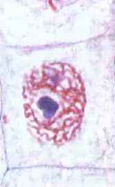
Rice. 67. Chromatin threads in the interphase of the cell life cycle
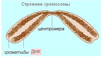
Most chromosomes in interphase are in the form of chromatin threads, which makes them practically invisible (Fig. 67). After reduplication, each chromosome consists of two DNA molecules that spiral, connect with proteins and take on distinct shapes. The two daughter DNA molecules are packaged separately and form sister chromatids (from the Greek chromium - color and eidos - appearance). Sister chromatids are held together and form one chromosome (Fig. 68). The site of cohesion between two sister chromatids is called a centromere (from the Latin centrum - middle and meros - part).
Rice. 68. Chromosome structure after DNA reduplication: 1 - centromere: 2 - chromosome arms; 3 - sister chromatids; 4 - DNA molecule: 5 - protein
It is possible to study the shape and size of chromosomes and determine their number in a cell only during division, when they are maximally spiralized, tightly packed, well stained and visible with a light microscope.
Chromosome set of cells. The cells of each organism contain a certain set of chromosomes, which is called a karyotype (from the Greek karyon - nucleus and typos - pattern, shape). Each type of organism has its own karyotype. Chromosomes of karyotypes differ in shape, size and set of genetic information. The chromosome set is strictly individual for each type of organism. Thus, the human karyotype consists of 23 pairs of chromosomes (Fig. 69), the fruit fly Drosophila has 4 pairs of chromosomes, and one of the wheat species has 14 pairs.
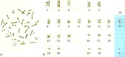
Rice. 69. Chromosome set of human cells: A - general photograph; B - 23 pairs of chromosomes
Studies of karyotypes of various organisms have shown that their cells may contain double and single sets of chromosomes.
A double set of chromosomes always consists of paired chromosomes that are identical in size, shape and nature of hereditary information. Paired chromosomes are called homologous (from the Greek homos - identical). Thus, all non-reproductive human cells contain 23 pairs of chromosomes, i.e. 46 chromosomes are presented in the form of 23 pairs. In Drosophila, 8 chromosomes form 4 pairs. Paired homologous chromosomes are very similar in appearance. Their centromeres are in the same places, and their genes are located in the same sequence.
Some cells may have a single set of chromosomes. For example, in the cells of lower plants - unicellular green algae - the set of chromosomes is single, while in higher plants and animals it is double. Animal sex cells also have a single set of chromosomes. In this case, there are no paired chromosomes, there are no homologous chromosomes, but there are non-homologous ones. Thus, human germ cells contain 23 chromosomes. Moreover, the chromosomal set of male and female germ cells differs in the 23rd chromosome. It resembles in shape the Latin letters X or Y. Spermatozoa may have an X or Y chromosome. Eggs always carry the X chromosome.
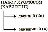
The chromosome set is usually denoted by the Latin letter p. The double set is accordingly denoted 2p, and the single set - p.
Exercises based on the material covered
- Define the life cycle of a cell (cell cycle).
- What is inferphase? What event is the main one in interphase? Justify your answer.
- How many DNA molecules does the chromosome consist of at the beginning of interphase and before cell division?
- How are the number and shape of chromosomes determined in different species of organisms?
- How does a double set of chromosomes differ from a single set?
- There are 44 chromosomes in the karyotype of a rabbit. How many chromosomes are in a rabbit's non-reproductive cells and how many are in the germ cells?
It was already mentioned above that in the cell nucleus, DNA molecules are located in special structures called chromosomes. Their research began over 100 years ago using a conventional light microscope. By the end of the 19th century, something had become clear about the behavior of chromosomes during cell division, and the idea was expressed about their participation in the transmission of heredity.
Chromosomes become visible in a microscope when a cell divides at a certain stage of the cell cycle called mitosis. Chromosomes in this state are compact rod-shaped structures of varying lengths with a fairly constant thickness; most chromosomes have a constriction that divides the chromosome into two arms. In the region of the constriction there is a structure important for the doubling of chromosomes, called centromere. When a cell divides during mitosis, the number of chromosomes doubles, as a result of which both newly formed cells are ultimately provided with the same standard set of chromosomes.
Only in 1956, for the first time, Y. Tio and A. Levan described the human chromosome set, determined the quantitative composition of chromosomes and gave their general morphological characteristics. In fact, these works marked the beginning of the study of the structure of the human genome. In humans, each cell of the body contains 46 chromosomes, the physical lengths of which range from 1.5 to 10 microns (Fig. 7).
Rice. 7. A microscopic view of the complete set of chromosomes contained in the nucleus of each individual human cell
Let us remind the reader that the set of chromosomes in all human cells (except for sex cells) is called diploid (double), since each chromosome is represented by two copies (23 pairs in total). Each human somatic cell (except red blood cells) contains 2 complete sets of chromosomes. Each single (haploid) set contains 23 chromosomes - 22 ordinary chromosomes (autosomes) and one sex chromosome - X or Y. Thus, the genome of each individual person consists of 23 pairs of giant DNA molecules distributed in different chromosomes, and if we say about the human genome in general (men and women), then the total number of such molecules is 24. This is the first basic information that was obtained about the human genome by analyzing chromosomes.
A study of the structure (size and shape) of human chromosomes showed that most of them resemble skittles in appearance, consisting of two thick parts (chromatids) and a thin constriction (centromeres) between them. The similarity with skittles and not with dumbbells is that the centromere is most often not located in the center of the chromosome, but is shifted to one of its ends. Chromosome sizes vary greatly, with the shortest chromosome being approximately ten times smaller than the longest. This is the second fundamentally important information about the structure of the human genome - the 24 DNA molecules that make it up have different sizes.
If you compare the number and size of chromosomes in humans and in other species of organisms, you can see huge differences. For example, a cow, whose genome size is approximately equal to the human genome, has 60 pairs of chromosomes. The clawed frog contains only 18 chromosomes, but even the smallest of them are larger than the largest human chromosomes. In birds, on the contrary, the number of chromosomes reaches 40 or more and they are all very small in size. Thus, the diversity of chromosomes in nature is very large.
Using light microscopy, the sizes of all human chromosomes were determined. Then all non-sex chromosomes were numbered in order of decreasing size - from 1 to 22. The sex chromosomes were not assigned a number, but were named X and Y. As more accurate subsequent studies showed, chromosome 21 actually turned out to be slightly smaller than 22, but the numbering of the chromosomes was not changed (so as not to cause confusion). The difference in the chromosome sets between men and women is that women have two sex X chromosomes (i.e., the chromosomes in all 23 pairs are the same), and in men a pair with the X chromosome is formed by the male sex chromosome - Y Each chromosome can be considered as a separate volume of a large twenty-four-volume collection of works called the Encyclopedia of Man.
Human germ cells, unlike the cells of the body of an adult organism (somatic cells), do not contain 2 sets of volumes of DNA text, but only one. Before conception, each individual chromosome (a separate volume in the Human Encyclopedia) of the father's sperm and mother's egg consists of different chapters of the DNA text of their parents mixed in different combinations. Any of the chromosomes we received from our father were formed in his testes shortly before we were conceived. Previously, in the entire history of mankind, such a chromosome had never existed. It was formed through the process of random mixing that occurs during division, gradually being formed from sections of the chromosomes of the ancestors on the father's side that unite with each other. The situation is the same with the chromosomes of eggs, except that they are formed in the body of our mother long before we are born (almost immediately after the birth of the mother herself).
In the zygote, which is formed as a result of the fusion of a sperm and an egg, the maternal and paternal genes are mixed and shuffled in different combinations. This occurs as a result of the fact that chromosomes do not remain unchanged over generations - they interact with their randomly encountered pair, exchanging material with it. This ongoing process is called recombination. And the next generation often gets a hybrid chromosome - part from the grandfather and part from the grandmother. Further in a series of generations, gene paths constantly intersect and diverge. As a result of the fusion of a unique egg with a unique sperm, a genome that is unique in all respects arises. And in this sense, we are all unique. Each human individual stores unique genetic information, consisting of a random combination of different gene variants.
A single gene can be viewed as a unit that continues to exist over numerous generations. And in this sense, the gene is immortal! There is even such an original point of view that it is not people themselves, but their genes that rule the world, and each specific living organism serves only as a temporary shelter for them. This controversial idea comes from Richard Dawkins, author of The Selfish Gene. In his opinion, genes are practically immortal, unlike the living organisms in which they exist. Some genes are tens or even hundreds of millions of years old. Genes, to use Dawkins' terminology, do whatever they can to survive. They adapt to heat and cold, choosing a better place for themselves, migrate with the help of humans and enter into new combinations. The man turned out to be a rather restless owner. For thousands of years, he has traveled extensively around the world, spreading his presence, influence and his stuff - genes. (The inquisitive reader can learn more about the ideas and argumentation of R. Dawkins in Appendix 1). This point of view is far from indisputable, and from further presentation it will become clear to us that genes are, first of all, not selfish, but workaholics. There are genes that are “guardians” of the genome, genes that are “janitors,” genes that are “cooks,” and genes that are “housekeepers.” By ensuring their existence, they ensure our existence.
Immediately after conception, the future person is just one cell (zygote), endowed with one initial DNA library containing 46 volumes. Among the 46 volumes, 23 are always received from the father and the other 23 from the mother. The texts of the 23 paternal and 23 maternal volumes, although very similar in general, nevertheless differ in details. For example, in the paternal volume No. 18 on page 253 there is a command sentence (in the form of a gene) that says that the child’s eyes should be brown, and in the same maternal volume on the same page it is also written about eye color, but according to This text color should be blue. The first indication is more strict (dominant) than the second, and as a result the child's eyes will have a brown color. The gene that dictates its rights is called dominant, and the one who cedes his rights - recessive. Only those people who have both maternal and paternal texts containing recessive genes that indicate blue-eyedness have blue eye color. Then the zygote divides into two cells, each of them divides again, and so on until billions of cells appear. The process of cell division is shown schematically in Fig. 8.
With each cell division, the volumes of DNA text contained in the libraries are copied exactly, with virtually no errors. The adult human body consists on average of 10 14 cells. For example, there are approximately 10 billion cells in the brain and liver, and 300 billion cells in the immune system. During a person's entire life, about 10 16 cell divisions occur in his body. The cellular composition of many organs is renewed several times over 70 years of life. And each of these cells contains the same 46 volumes of DNA text.
At the end of the 60s of the 20th century, an important breakthrough was made in the study of chromosomes. It was only due to the fact that they began to use a special contrast agent for their coloring - akrichine mustard, and then other compounds similar to it. This staining made it possible to identify a large number of different substructures inside chromosomes that were not visible under a microscope without staining. After staining the chromosomes with a specific Giemsa-Romanovsky dye, they look like zebras: transverse light and dark stripes of varying color intensity are visible along the entire length.
Rice. 8. The main stages of the cell cycle leading to cell division
These bands are called chromosomal G segments or bands (Fig. 9). The pattern of segmentation varies greatly among different chromosomes, but the arrangement of chromosomal segments is constant on each chromosome in all types of human cells.
The nature of the stripes revealed by staining is not yet completely clear. It has now only been established that regions of chromosomes corresponding to dark bands (called R-bands) replicate earlier than light regions (called G-bands). Thus, the banding of chromosomes most likely still has some meaning that is not yet fully understood.
Staining chromosomes greatly facilitated their identification, and subsequently contributed to determining the location of genes on them (gene mapping).
Rice. 9. Specific chromosomal G-segments identified by staining human chromosomes, and the system of their designation according to the decision of the international conference in Paris in 1971. The numbers below the chromosomes indicate their numbers. X and Y - sex chromosomes, p - short arm, q - long arm of chromosomes
Although the detailed processes occurring during staining are not yet completely clear, it is obvious that the coloring pattern depends on such a parameter as the increased or decreased content of AT or GC pairs in individual chromosome bands. And this is another general information about the genome - it is not homogeneous, it contains regions enriched with certain nucleotide pairs.
This, in particular, may be due to the repeatability of certain types of DNA nucleotide sequences in certain regions.
Differential coloring of chromosomes has found wide application for the detection and identification of small individual changes in the genome of a particular person ( polymorphism), in particular, leading to various pathologies. An example of this is the discovery of the so-called Philadelphia chromosome, which is found in patients with chronic myeloid leukemia. Using chromosome staining, it has been established that in patients with this disease, a certain fragment disappears on chromosome 21 and appears at the end of the long arm of chromosome 9 (fragment transfer or translocation, abbreviated t). Geneticists designate such an event as t (9; 21). Thus, chromosomal analysis indicates that different DNA molecules can exchange separate sections with each other, resulting in the formation of “hybrids” in the genome, consisting of DNA molecules of different chromosomes. Analysis of the already studied properties of chromosomes made it possible to form an idea of the polymorphism of the human genome.
To determine the localization of individual genes on chromosomes (that is, gene mapping), a whole arsenal of special methods, often very complex in design and execution, is used. One of the main ones is molecular hybridization (formation of a hybrid) of a gene or its fragment with chromosome preparations fixed on a solid support, isolated from cells in pure form (this is called hybridization in situ). The essence of the hybridization method in situ consists in the interaction (hybridization) between denatured (unbraided) DNA strands in chromosomes and complementary nucleotide sequences of chromosomes added to the preparation, individual single-stranded DNA or RNA (they are called probes). If there is complementarity between one of the chromosomal DNA strands and the probe, fairly stable molecular hybrids are formed between them. The probes are pre-labeled using different labels (radioactive, fluorescent, etc.). Places of hybrid formation on chromosomes are identified by the position of these marks on chromosome preparations. Thus, even before the advent of genetic engineering methods and DNA sequencing, they found out, for example, the location in the human genome of genes encoding large and small ribosomal RNAs (rRNAs). The genes of the former turned out to be localized in five different human chromosomes (13, 14, 15, 21 and 22), while the bulk of small rRNA genes ( 5S RNA) is concentrated in one place on the long arm of chromosome 1.
An example of a picture obtained by hybridization of fluorescent dye-labeled gene probes is shown in Fig. 10 on the colored insert.
Rice. 10. Hybridization of human chromosomes with gene probes labeled with red and green fluorescent dyes. The arrows indicate the location of the corresponding genes at the ends of two different chromosomes (the top right shows an enlargement of the picture of hybridizing chromosomes).
Genes located on the same chromosome are defined as linked genes. If genes are located on different chromosomes, they are inherited independently (independent segregation). When genes are on the same chromosome (that is, linked), they are incapable of independent segregation. Occasionally, various changes in chromosomes can occur in germ cells as a result of recombination processes between homologous chromosomes. One of these processes is called crossing over. Because of crossing over, linkage between genes of the same group is never complete. The closer the linked genes are to each other, the less likely it is that the location of such genes will change in children compared to their parents. Measuring the frequency of recombination (crossing over) is used to establish the linear order of genes on a chromosome within a linkage group. Thus, when mapping chromosomes, it is initially determined whether these genes are located on the same chromosome, without specifying which one. After at least one of the genes of a given linkage group is localized on a specific chromosome (for example, using hybridization in situ), it becomes clear that all other genes of this linkage group are located on the same chromosome.
The first example of the connection of genes with certain chromosomes can be the detection of the linkage of certain heritable traits with sex chromosomes. To prove the localization of a gene on the male sex Y chromosome, it is enough to show that this trait is always found only in men and is never found in women. The linkage group of the female X chromosome is uniquely characterized by the absence of heritable characteristics transmitted from father to son and the inheritance of maternal characteristics.
Particularly important for studying the human genome in the early stages of its research was a method called somatic cell hybridization. When human somatic (non-reproductive) cells are mixed with cells of other animal species (most often, mice or Chinese hamster cells were used for this purpose), fusion of their nuclei (hybridization) can occur in the presence of certain agents. When such hybrid cells reproduce, some chromosomes are lost. By a happy accident for the experimenters, in human-mouse hybrid cells, most of the human chromosomes are lost. Next, hybrids are selected in which only one human chromosome remains. Studies of such hybrids have made it possible to associate some biochemical characteristics characteristic of human cells with certain human chromosomes. Gradually, through the use of selective media, they learned to achieve the preservation or loss of individual human chromosomes carrying certain genes. The selection scheme, although not very simple at first glance, showed itself quite well in the experiment. Thus, they came up with a special selective medium in which only those cells in which the enzyme thymidine kinase is synthesized can survive. If for hybridization with human cells we take as a partner mutant mouse cells that do not synthesize thymidine kinase, then only those hybrids that contain human chromosomes with the thymidine kinase gene will survive. In this way, for the first time, it was possible to establish the localization of the thymidine kinase gene on human chromosome 17.
Despite the fact that the study of the human genome at the chromosome level provided a number of important characteristics, they were the most general and provided relatively little for a complete understanding of the structure and functioning of the genetic apparatus of human cells.
| |
We said that somatic cells contain a double, diploid set of chromosomes, and mature sex cells contain a single, haploid set. A diploid set of chromosomes is also present in unripe germ cells. A halving of the number of chromosomes and, accordingly, DNA, referred to as their reduction, occurs in the process of gametogenesis, that is, the development of germ cells - gametes. Reducing by half the number of chromosomes in gametes is a preparation for future fertilization, in which their diploid set is restored due to the combination of the haploid chromosome sets of the sperm and egg.
Gametogenesis occurs in the gonads: in the testes in the male body and in the ovaries in the female. Accordingly, it is called spermatogenesis and oogenesis. Common to spermato- and oogenesis are the first 3 periods of gametogenesis: reproduction, growth and maturation. Developing male germ cells undergo an additional fourth period - formation (Fig. 26).
In the first period of gametogenesis, germ cells intensively divide mitotically and their number increases. In this period, the germ cells are called spermato- and oogonia, respectively. For simplicity, the diagram shows the case when the haploid set includes only 3 chromosomes, one of which is sexual - gonosome, or heterochromosome (in Greek, "heteros" means different) and two non-sexual - autosomes. In the cells, one haploid set is blackened, the second is indicated by contour lines. Homologous, unambiguous autosomes of both sets are drawn in the same shape and size (this is the longest line and circle). Heterochromosomes are depicted differently - with a straight line (X chromosome) and a curved line of the same length (Y chromosome). Chromosomes lie separately in the cell.
Sex cells that have entered the growth period are designated as spermato- and oocytes of the first order. They increase in size, especially oocytes, and their nuclear apparatus undergoes restructuring. Homologous chromosomes lie parallel to each other, forming bivalents, the number of which is equal to the number of chromosomes in the haploid set. Each of the two chromosomes of the bivalent, in turn, is a pair structure - a dyad, since it consists of two sister chromatids. When the gap between these chromatids becomes well defined, the bivalents already look like tetrads. The number of tetrads corresponds to the haploid number of chromosomes. The total number of detectable chromatids - future chromosomes of mature germ cells - is tetraploid. The amount of DNA in spermato- and oocytes before the maturation period is also tetraploid.
Then comes a period of maturation, which is characterized by meiosis (“meiosis” means reduction) - two rapidly successive divisions of germ cells, during which chromosome reduction occurs. Male germ cells that have completed the first division of maturation are called spermatocytes of the second order, or prespermatids, and the corresponding female germ cells are called oocytes of the second order. After the second division of maturation, prespermatids become operamatids, oocytes of the second order become mature eggs. The first division of maturation is reduction. During this division, whole chromosomes - dyads - are distributed between the daughter cells and the chromosome set becomes haploid.The second division of maturation is called equalizing, equational, since the halves of the chromosomes (dyads), essentially their chromatids, diverge between the daughter cells.
The interphase between the first and second divisions of maturation can be very short or even absent altogether, since here at this time neither DNA replication nor doubling of the number of chromosomes occurs in the cells. Division begins again, now of two cells, and each of the grandchild cells receives one chromosome from the dyad. Thus, each of these four cells, resulting from 2 divisions of maturation, receives one of the elements of the tetrad. The haploid number of tetrads corresponds to the haploid number in the chromosome set of cells that have undergone meiosis. In the same way, the amount of DNA that is tetraploid at the beginning of meiosis (in the oocyte and spermatocyte of the first order) after division into 4 parts becomes haploid at the end of it.
During spermatogenesis, from each spermatogonia that has entered the growth period, 4 full-fledged spermatids are obtained as a result of maturation divisions. During oogenesis, the second-order oocyte emerging from a first-order oocyte retains almost the entire size of the mother cell. The second daughter cell receives half of the mother's chromosomal material and only an insignificant part of its cytoplasm. This small cell is called the reduction body. A similar picture is repeated during the second division of maturation - a second-order oocyte gives rise to an egg approximately equal in size and a second reduction body. At the same time, the first reduction body also divides into two. As a result, from one oogonia, which has passed from the period of reproduction to the period of growth, and then into the period of maturation, only one mature egg is formed. This is an advantageous adaptation for procreation - the entire supply of nutrients accumulated by the first-order oocyte during the growth period, necessary to ensure the initial stages of development of the future embryo, remains in the egg.
During the formation period, the spermatid of a typical cell is transformed into a spermatozoon, which has a very complex structure, ensuring its role as an active, mobile partner in the act of fertilization. All components of the spermatid participate in this restructuring.
First of all, its centrioles are located one after another, thus determining the long axis of the future sperm (Fig. 27A). Among the many small proacrosomal granules formed in the spermatocyte - in the center of its Golgi apparatus, which now moves to the anterior end of the cell (Fig. 27B-3), one large acrosomal (“acron” means tip) granule appears, which then lies at the nucleus in the place of its future head pole (Fig. 27B-9). The lamellar complex is reduced, giving rise to an acroblast vesicle enveloping the acrosomal granule. The body of the spermatid begins to gradually lengthen, and the cell nucleus becomes more and more dense. It is located at the anterior end of the developing sperm. The proximal centriole lies behind the nucleus, and the distal one forms a flagellum, like a kinetosome. Then it is divided into two parts, and the rear of them takes the shape of a ring (Fig. 27B-8) and moves away from the front, sliding along the growing flagellum - the future axial filament of the sperm tail. The ring lingers at the rear edge of the cell. By this time, the mitochondria are already mostly accumulated near the axial filament. The acroblast, growing, moves in the form of a cap onto the front part of the nucleus.
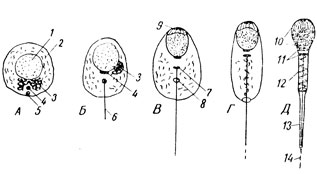
At the end of the formation period, differentiation into sections is well expressed in the sperm: the head, which is basically a flattened and very compact nucleus, covered in front by a sheath, under which the acrosome lies at the very edge; a neck formed by centrioles; intermediate, connecting section and tail. All mitochondria are concentrated in the intermediate section, which spiral around the axial filament. The distal boundary of the intermediate section is the closing centriole ring. In the cytoplasm there are relatively many substances (glycogen, lipids), due to the breakdown of which the sperm partially receives energy for movement. In the tail, when examined under an optical microscope, two sections are distinguished - the main one, covered with cytoplasm, and the terminal one, “bare”, consisting only of the tail filament.
Using an electron microscope, it was established that the thin cap covering the sperm nucleus is a flattened cistern. Its inner membrane is adjacent to the nucleus, and its outer membrane is adjacent to the plasmalemma. At the back of the sperm head, the plasmalemma directly covers the nucleus. The core itself is very densely filled with intertwining threads 40 Å thick, which are DNP (nucleohistone) molecules. Chemical analysis of condensed nuclear chromatin shows that it consists of approximately half DNA and half protein. The centrioles located in the neck region have a structure typical of these organelles. The smaller of them, adjacent to the back of the nucleus in its middle part, has the appearance of a cylinder formed by 9 pairs of tubes. The distal centriole is more developed. It, like a kinetochore, is in connection with 9 pairs of filament edges of the flagellum, which contains 2 more central ones, like all cilia and flagella in general.
The mitochondria, densely lying in the intermediate section around the axial filament, form approximately 14 turns of the spiral. Apparently, first of all, they provide energy to the contractile elements of the sperm. In the main section of the tail, 9 pairs of axial filaments are surrounded by ring fibrils, which are held together by two longitudinal thick strands. In the terminal section of the tail, there are no ring fibrils; the bundle of axial fibrils forming the axial filament is enclosed in a homogeneous mass and covered on the surface by a plasmalemma. Thus, here we encounter a structure characteristic of all cilia and flagella.
The total length of sperm in humans reaches approximately 60 mk. It moves actively at a speed of 3.5 mm per minute. At the same time, it rotates around its axis clockwise, making one full revolution in 15 minutes. Its ability to move depends on the pH and other properties of the environment. The lifespan of sperm in the vagina is only 1 hour; in other female genital tracts it lasts several days. Along the female genital tract, the sperm moves passively (as a result of muscular contractions of the walls of the uterus and oviducts), reaching the upper third of the oviducts, where fertilization occurs. Thus, he does not have to waste energy, from his generally insignificant reserve, to overcome such a long path for him.
A mature egg cell has a spherical shape; its diameter in humans is 135 mk. It is always covered with a microscopically visible membrane and its structure differs from somatic cells, mainly in two ways. Firstly, in it the nuclear-cytoplasmic ratio is more or less sharply shifted in favor of the cytoplasm, which is explained by the accumulation in the body of the oocyte during the period of growth of nutrients for the needs of the future embryo. Secondly, it lacks a cell center, which disappears during the same period of growth. The cellular center introduces sperm into the egg during fertilization, and after this, the mitotic division of the zygote begins - an organism located at the unicellular stage of ontogenesis.
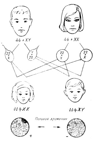
Let us briefly touch upon the issue of sex determination, which arises after fertilization of the organism. As it turned out, this occurs at the moment of conception, that is, fertilization, and is caused by the combination of heterochromosomes in the zygote. Let us turn to the diagram (Fig. 26) and consider the chromosome sets in developing female and male germ cells. All oogonia have two X chromosomes. It follows that the haploid set of chromosomes of each mature egg will necessarily include one of the X chromosomes. In spermatogonia, among the two heterochromosomes, there is one X chromosome and one Y chromosome. Therefore, the sperm must be different - half with an X chromosome and half with a Y chromosome.
In mammals and humans, when two X chromosomes are combined in a zygote, a female organism develops; in the presence of XY chromosomes, a male organism develops. Thus, the sex of the unborn child will depend on which of the father’s sperm the mother’s egg connects with (Fig. 28). The human haploid set contains one or another of the heterochromosomes (sex chromosomes - gonosomes) and 22 autosomes (non-sex chromosomes). There should be 46 chromosomes in the zygote, embryonic, somatic and immature germ cells of a human - 44+XY in the male body and 44+XX in the female. Only after the first meiotic division in the maturing germ cells is the number of chromosomes reduced to 23. For normal development to occur, it is necessary to have a diploid set of chromosomes without any deviations, both quantitative and structural.
In 1949, Barr established that in female mammals and women, the X chromosomes in the diploid set behave differently - one of them, after cell division, despirals, like autosomes, and becomes indistinguishable in the interphase nucleus, the other remains highly spiraled, like heterochromatic chromomeres . This heterochromatic X chromosome, clearly visible in the nucleus in the form of a dark body in the usual staining of preparations, is called sex chromatin. In genetic experiments to identify Barr bodies (sex chromatin bodies), it is easiest to examine blood lymphocytes or desquamated epithelial cells. It was found that either of the two X chromosomes could become inactive.
In rare cases, during oogenesis, the X chromosomes do not separate during meiosis. As a result, eggs that deviate from the norm may be formed: instead of one X chromosome, there may be two, or there may be no X chromosome at all. In the latter case, during fertilization, two types of zygotes can arise, containing in the diploid set either one Y chromosome or one X chromosome introduced into the egg during fertilization by a sperm. A zygote with one Y chromosome is generally not viable and dies. A zygote with one X chromosome in humans has 45 chromosomes: 44+XO. With this combination of chromosomes, the zygote develops into an inferior woman of small stature with rudimentary ovaries and, as a consequence, the absence of secondary sexual characteristics. This pathology is known as Turner syndrome. The only X chromosome in this case is despiralized, and therefore sex chromatin is not detected in somatic cells in such girls.
The result of non-disjunction of X chromosomes during meiosis will also be deviations in the set of the opposite nature, namely the presence of three X chromosomes or two X chromosomes and one Y chromosome in the zygote. A female organism with three X chromosomes is referred to as a “superfemale” or “superwoman” (for humans). However, such an individual is called a “superwoman” only conditionally, based on the supernumerary number of X chromosomes. In reality, with a set of chromosomes 44 + XXX, there is underdevelopment of the ovaries and therefore often a loss of fertility. Interestingly, “superwomen” with four X chromosomes (chromosomal set 44 + XXXX) are fertile, but have reduced mental development. When analyzing the sex chromatin of somatic cells in women with XXX or XXXX chromosomes, 2 or 3 Barr bodies are found in the nuclei, respectively. Thus, in these cases, only one of all X chromosomes is despiralized and active in interphase nuclei.
Zygotes with a chromosome set of type 44+XXY develop in men suffering from Klinefelter syndrome - mental retardation and underdevelopment of the testis, leading to infertility. Their somatic cells have sex chromatin and contain 1 Barr body. The second X, as in normal men, is despiraled in interphase. Similar developmental defects are observed with a larger number of X chromosomes combined with the Y chromosome, namely with types XXXY, XXXXY and XXXXXY. The presence of the Y chromosome in the set determines the development of a male individual, but an inferior one. The number of Barr bodies in the interphase nuclei of somatic cells is one less than the number of X chromosomes in the set.
Relatively recently, another chromosomal anomaly was discovered - type XYY. Animals “super males” with an extra Y chromosome are distinguished by great strength and aggressiveness. Men with two Y chromosomes are tall (above 180 cm), have great physical strength, but have reduced mental abilities. As in normal men, the interphase nuclei of their somatic cells do not contain sex chromatin.
Scientists have found that the inheritance of some diseases is associated with genes located on the sex chromosomes. For example, congenital color vision disorders (a disease formerly called color blindness), in general blindness caused by atrophy of the optic nerve, hemophilia (pathological difficulty stopping bleeding) are transmitted through the X chromosome.
The phenomenon when there are extra chromosomes in a set or some of the chromosomes are missing is called aneuploidy. The presence of one extra chromosome is referred to as trisomy; if there are two such extra chromosomes, then it is double trisomy. If one chromosome is missing, it is called monosomy.
An example of disorders associated with an increase in the number of autosomes is the most famous trisomy - one of the smallest chromosomes - the 21st. The presence of 21 chromosomes in the chromosome set instead of two or three occurs in Down syndrome - one of the forms of mental retardation, combined with delayed and impaired physical development, and sometimes with the presence of certain deformities (the appearance of patients is very similar and is characterized by a small skull , flat back of the head, slanted eyes, wide sunken bridge of the nose, half-open mouth, deformed ears). The genitals are underdeveloped, and secondary sexual characteristics are poorly expressed. Half of children with Down syndrome do not live beyond 2 years of age.
Many diseases have been discovered that are caused by genes located on different autosomes. Among them can be named inherited mental illnesses, such as schizophrenia and epilepsy.
All kinds of hereditary human diseases caused by various genotype disorders are the subject of study of the currently intensively developed branch of cytogenetics - medical genetics.
Chromatid- (from the Greek chroma - color, paint and eidos - appearance) - a structural element of the chromosome, formed in the interphase of the cell nucleus as a result of chromosome doubling (replication).
Chromatid- nucleoprotein thread, half of a double chromosome. A chromosome can be single (from one chromatid) or double (from two chromatids).
The function of chromosomes is to store hereditary information. Chromosomes are located in the nucleus of the cell and are the main components of the nucleus.
Chromatid is any of the two copies of a DNA molecule that together make up a replicated chromosome and are connected by their centromeres. This term is used as long as the centromeres remain in contact. After chromosomes separate during anaphase of mitosis or meiosis, the strands are called daughter chromosomes. In other words, chromatids are halves of replicated chromosomes. The chemical composition of chromosomes is 50% DNA and 50% protein.
Humans typically have 23 pairs of homologous chromosomes in each cell (N = 23). However, the number of chromatids will be a multiple of 23 and may be 4N, 2N or 1N. The number does not refer to the haploid or diploid set, it refers to the number of chromatids in each cell, as a multiple of the haploid set of chromosomes in the body.
- 4N. In a cell with 4N chromatids, there are 23 pairs of chromosomes (46 chromosomes), and each chromosome has two chromatids. Thus, there are 92 chromatids in each cell (4N).
- 2N. Immediately after mitosis, during which the cell divides in half, 23 pairs of chromosomes (46 chromatids) appear. However, a chromosome has only one chromatid. Thus, there are 46 chromatids in total (2xN). On the other hand, a haploid cell with two chromatids per chromosome also has 46 chromatids. However, this does not happen in humans.
- 1N. Immediately after meiosis, each cell, called gamete, has only half the amount of chromosomes (23 chromosomes). In addition, each chromosome has only one chromatid. Thus, there are 23 chromatids in total (1xN).
A single chromosome turns into a double chromosome during the process of replication (doubling of DNA). A double chromosome turns into two single chromosomes (chromatids become daughter chromosomes) after the separation of the centromere connecting them (in anaphase of mitosis and anaphase II of meiosis). That is chromatids become chromosomes after the centromere connecting them divides.
Chromosome constrictions:
Chromosomes- the main carriers of genetic material, ensuring its transmission from generation to generation.
During the period between cell divisions (in the interphase of mitosis), chromosomes are invisible in a light microscope and are represented by untwisted (despiralized) chromatin threads. During this period, an important genetic event occurs - DNA replication and the doubling of chromosomes based on it. As long as the two resulting copies are held together by the centromere, they are called sister chromatids. With the onset of cell division, chromosomes spiral and become denser. Under a light microscope, it becomes clear that they consist of two chromatids. During mitosis, chromatids separate and become independent chromosomes. Thus, during the cell cycle, the structure of chromosomes undergoes changes.
Each chromosome is individual, i.e. characterized only by its size, shape and position of the centromere. In the body cells of organisms that reproduce sexually, any chromosome is represented by two copies, or homologues. When germ cells are formed in meiosis, each of them receives one of two homologous chromosomes. During fertilization, the pairing of homologous chromosomes is restored: one chromosome of each pair is paternal, the other is maternal.
The set of characteristics of a chromosome set (the number of chromosomes, their size and shape) is constant for cells of each species and is called its karyotype. A karyotype distinguishes between a pair of sex chromosomes that determine the sex of an organism, and all other chromosomes are autosomes. The study of the behavior of chromosomes in mitosis and meiosis, as well as the role of chromosomes, especially sex chromosomes, in the transmission of traits from one generation to another led to the creation of the chromosomal theory of heredity at the beginning of the 20th century. A chromosome is often referred to as the genetic material of bacteria and viruses., although its structure differs from the chromosomes of eukaryotic organisms.
Sets of chromosomes
A single (haploid) set of chromosomes is characteristic of germ cells ( gametes), double (diploid) - for somatic cells.
- A single set turns into a double set at the moment of fertilization (fusion of sperm and egg).
- The double set turns into a single set at the moment of independent divergence of double chromosomes in anaphase I of meiosis.
The human double set contains 46 chromosomes.
Problems on the number of chromosomes:
- If there are 12 chromosomes in a reproductive cell, then in a fertilized egg ( zygote) and in somatic cells(liver, skin, muscles, etc.) - 24 chromosomes each.
- And vice versa: if there are 36 chromosomes in the nucleus of a somatic cell (intestine, brain, etc.), then after meiosis 18 remain in the eggs and sperm, and when the eggs and sperm merge, there will again be 36 in the zygote nucleus.
Where can you find lists of lawyers in Russia on the Internet?
Information about the bar chambers of the constituent entities of the Russian Federation (number of members, presidents of the bar chambers, addresses, telephone numbers, websites), lists of legal bureaus, offices, boards, lists of lawyers included in the registers of the constituent entities of the Russian Federation, as well as information about a specific lawyer or check whether a person is a lawyer, whether his powers have been terminated can be found on the websites: Register of lawyers
What does the name Galina mean?
Galina - from the Greek word galene, which means calm, serenity. Derived forms: Alya, Gala, Galinka, Galyunya, Galyusya, Galyukha, Galyusha, Galya, Pebble, Galenka, Galonka, Galka, Galochka, Galchonok, Galchonochek. Name days: February 23, March 23, April 29. (Attention! Further
In what year did Prada release its first fragrance?
Prada is a well-known Italian private company specializing in the production of fashionable clothing, shoes and accessories, which owns the fashion house and trademark of the same name. Prada's style is recognizable, it is laconic, strict and elegant. The history of the brand began in Milan, in 1913, when Mario Prada -
How is botulism treated?
What is botulism?Botulism is an acute infectious disease that occurs as a result of ingestion of food, water or aerosols contaminated with the spore-forming bacillus Clostridium botulinum. Manifests itself as a violation of contractions of striated and smooth muscles. Botulinum toxin is absorbed from the gastrointestinal tract or lungs
What are the advantages and disadvantages of LED lamps
LED lamps or LED lamps use LEDs as a light source and are used for household, industrial and street lighting. The LED lamp is one of the most environmentally friendly light sources. The LED glow principle allows the use of safe components in the production and operation of the lamp itself. I don't use LED lamps
What to give a teacher on Teacher's Day
Teacher's Day is a professional holiday for education workers. History of the holiday The historical prerequisite for the establishment of Teacher's Day was the Special Intergovernmental Conference on the Status of Teachers held on October 5, 1966 in Paris.
Attention! All treatments must be carried out after consultation with a professional doctor and under his constant supervision. The patient himself is responsible for the consequences of using the information provided. Gastritis is inflammation of the mucous (inner) lining of the stomach wall. When inflammation spreads to the duodenum, gastroduodenitis is formed. There are two types
What order does the light hawk belong to?
The light hawk (lat. Accipiter novaehollandiae) is a bird of prey of the hawk family, common in the forests of northern and eastern Australia. Kingdom: Animals Type: Chordata Class: Birds Order: Falconiformes Family: Accipitridae Subfamily: Accipites Genus: True hawks Species: Light hawk Light hawk &md
When was International Music Day established?
International Music Day is celebrated on October 1st. International Music Day was established on October 1, 1975 by decision of UNESCO. One of the initiators of the establishment of International Music Day was Dmitry Shostakovich, a classical composer of the 20th century. It is celebrated annually all over the world with large concert programs with the participation of the best artists and artistic groups. In e
How Christmas is celebrated in Denmark
Christmas is celebrated all over the world, and many countries have their own traditions and customs associated with its celebration. Since ancient times, the Day of the Nativity of Christ has been ranked among the great holidays by the Church, according to the Gospel, which depicts this event as great and wonderful: “I proclaim to you,” says the angel to the Bethlehem shepherds, “a great
Which famous people died on December 27
December 27 is the 361st day of the year (362nd in leap years) in the Gregorian calendar. There are 4 days left until the end of the year. Holidays Russia, Rescuer Day; Day of the Apostle and Evangelist John (holiday in Catholic and Protestant denominations). Name day Firs, Leonid, Gennady, Hilarion, Nikolai. Events of December 27, 6th century
1. Somatic cells, as opposed to germ cells
- incapable of dividing
- contain n chromosomes
- contain 2n chromosomes
Somatic cells– cells of the body of an animal or plant (i.e. non-reproductive cells). In somatic cells (body cells), the number of chromosomes is usually twice as large as in mature germ cells. This is explained by the fact that during fertilization, half of the chromosomes come from the mother’s body (in the egg) and half from the father’s (in the sperm), i.e. In the nucleus of a somatic cell, all chromosomes are paired. Moreover, the chromosomes of each pair are different from other chromosomes. Such chromosomes, identical in shape and size, carrying identical genes, are called homologous. One of the homologous chromosomes is a copy of the maternal chromosome, and the other is a copy of the paternal chromosome. The chromosome set, represented by paired chromosomes, is called double or diploid, and is designated 2n.
2. Human cells are different from chimpanzee cells
- presence of ribosomes
- number of chromosomes
- lack of DNA
Human cells differ from chimpanzee cells in the number of chromosomes: humans have 23 pairs of chromosomes, great apes have 24. Chimpanzees are our closest relative with almost the same karyotype as ours (the pygmy chimpanzee is especially close to us in terms of chromosomes).
The chromosome set of a somatic cell, in which each chromosome has a pair - double or diploid ( 2n). The amount of DNA corresponding to the diploid set of chromosomes is − 2s. From each pair of homologous chromosomes, only one gets into the germ cells, so the chromosome set of gametes is single or haploid ( 1n).
3. Sphagnum spores are special cells that carry out
- asexual reproduction
- sexual reproduction
- fragmentation
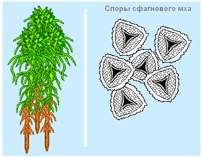 Sphagnum can reproduce both by spores and vegetatively.
Reproduction by spores is the main method for the dispersal of sphagnum over long distances - new areas or areas damaged by fire or economic activity. In the upper part of the plant, boxes on stalks ripen, in which spores are formed.
To form a plant from a spore, it is necessary that it fall on suitable soil - moist peat. Larger spores have a greater supply of nutrients and therefore a better chance of waiting for the right conditions.
4. Specialized cells with the help of which sexual reproduction is carried out are called
- disputes
- blastomeres
- oocytes
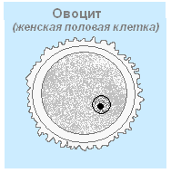
The process of development of female germ cells - eggs from the primordia of the epithelium (oogenic tissue). Ontogenesis occurs in the ovaries in three phases: reproduction, growth and maturation. The first phase - reproduction - cells of the diploid tissue of the rudimentary epithelium divide repeatedly through mitosis, forming diploid cells oocytes (oocytes) first order.
5. The picture shows 
- chromosome
- chromatid
- centriole
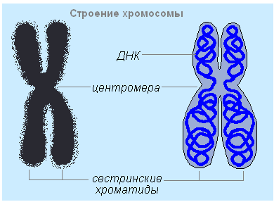 Chromosomes– the most important structures of the nucleus. During the period between cell divisions (interphase), chromosomes are not visible. They can be in two states: spiralized - short and dense, clearly visible in a light microscope; despiralized (untwisted) - long and thin, called chromatin (colored substance). There are no differences in the chemical composition of chromosomes and chromatin; These are DNA molecules and proteins (proteins) that together form a nucleotide. In the metaphase of a dividing cell, it is clearly visible that each chromosome consists of two longitudinally symmetrical halves - chromatids. In specialized cells, chromosomes are usually single-chromatid. The chromosome has a primary constriction on which the centromere is located; the constriction divides the chromosome into two arms of equal or different lengths. The centromere connects both chromatids and serves as the attachment point for the spindle filaments of cell division. The main function of chromosomes is the storage and transmission of hereditary information, the carrier of which is the DNA molecule - genes - which are responsible for each specific trait or property of the organism. Hereditary information is transmitted from cell to cell by doubling the DNA molecule (replication), transcription and translation.
6. The chromosome contains a molecule
- glucose
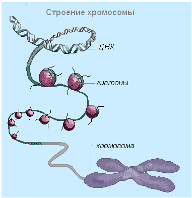
A chromosome consists of DNA and squirrel. A complex of proteins bound to DNA forms chromatin. Proteins play an important role in packaging DNA molecules in the nucleus. Before cell division, DNA is tightly coiled to form chromosomes, and nuclear proteins - histones - are necessary for the correct folding of DNA, as a result of which its volume decreases many times. Each chromosome is formed by one molecule DNA.
7. During sexual reproduction, each species maintains a constant number of chromosomes from generation to generation due to
- mitosis
- meiosis
- gametogenesis
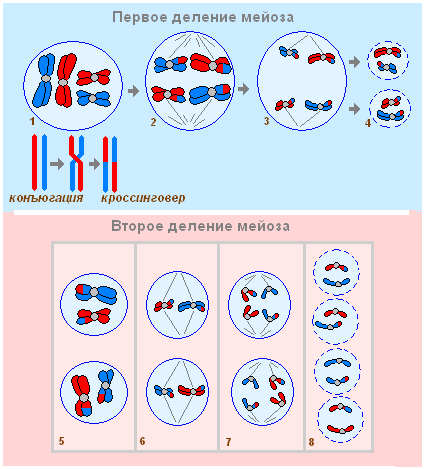
Meiosis is a special type of cell division, as a result of which daughter cells become haploid. Meiosis consists of two divisions occurring one after the other; DNA reduplication occurs only once. All necessary substances and energy (ATP) accumulate in interphase I, interphase II is practically absent.
First division of meiosis.
1. Prophase I - spiralization of chromosomes with the formation of bivalents (a structure consisting of two chromosomes and four chromatids), chromosome conjugation - bringing together two homologous chromosomes along the entire length and crossing over - exchange of sections of homologous chromosomes, disappearance of the nucleolus, formation of a spindle, destruction of the nuclear membrane. (2n 2chr 4c) – 1n bivalent
2. Metaphase I – arrangement of pairs (bivalents) of homologous chromosomes at the equator of the cell. (2n 2chr 4c) – 1n bivalent
3. Anaphase I – divergence of homologous chromosomes, consisting of two chromatids, to opposite poles of the cell (the number of chromosomes is reduced). (1n 2chr 2c)– at each pole of the cell.
4. Telophase I – formation of nuclei, division of the cytoplasm – formation of two daughter cells. (1n 2chr 2c)
Second division of meiosis.
5. Prophase II – slight spiralization of chromosomes, formation of a spindle, destruction of the nuclear membrane. (1n 2chr 2c)
6. Metaphase II - ordered arrangement of chromosomes, consisting of two chromatids, at the equator of the cell. (1n 2chr 2c)
7. Anaphase II – separation of daughter chromatids to opposite poles of the cell. (1n 1chr 1c)- at each pole.
8. Telophase II - disappearance of the spindle, despiralization of chromosomes, formation of nuclear membranes, division of the cytoplasm, formation of new cell membranes, formation of four daughter cells. (1n 1chr 1c)– in each formed nucleus.
Meiosis serves as the basis for sexual reproduction and combinative variability.
8. Chromosomes include
- ATP and mRNA
- RNA and protein
- DNA and protein
In an interphase cell, chromatin has the form of fine-grained thread-like structures consisting of molecules DNA and protein(nucleoprotein) sheath. In dividing cells, chromatin structures spiral and form chromosomes. A chromosome consists of two chromatids, and after nuclear division it becomes single chromatid.
The DNA molecule in chromosomes is closely associated with two classes of proteins - histones (basic proteins) and non-histones (acidic proteins). Histones are small proteins with a high content of charged amino acids (lysine and arginine) they are necessary for the assembly and packaging of threads DNA into chromosomes.
9. Chromosome doubling occurs
- anaphase
- interphase
- prophase
Interphase – preparation of the cell for division, consists of three periods
Designations of genetic material.
1n– haploid set of chromosomes
2n– diploid set of chromosomes
chr- quantity
chromatids on one chromosome
c– number of chromatids in a set of chromosomes
2c– the amount of DNA corresponding to the diploid set of chromosomes
10. Somatic cells are formed as a result
- mitosis
- fertilization
- oogenesis
11. Human leukocytes contain... chromosome(s)
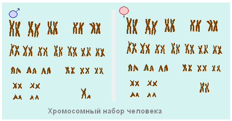
Human chromosomes contain genes (46 units), forming 23 pairs. One pair of this set determines the gender of a person. A woman's set of chromosomes contains two X chromosomes, a man's - one X and one Y. All other cells of the human body contain twice as many as sperm and eggs.
12. The separation of sister chromatids occurs in ... mitosis
- anaphase
- metaphase
- prophase
IN anaphase(4) sister chromatids are separated under the action of the spindle: first in the centromere region, and then along the entire length. From this moment on, they become independent chromosomes. The spindle threads stretch them to different poles. Thus, due to the identity of the daughter chromatids, the two poles of the cell have the same genetic material: the same as what was in the cell before the start of mitosis.
Amount of genetic material 2n 1chr 2c at each pole of the cell.
13. Zygote, as opposed to gamete
- formed during mitosis
- has 2n chromosomes
- formed during the process of meiosis
A zygote is the first cell of a new organism, which contains in the nucleus the genetic material of the sperm and egg, i.e. the diploid set of chromosomes is restored: =n + n = 2n.
14. New somatic cells in the body of an earthworm are formed as a result
- mitosis
- gametogenesis
- meiosis
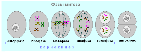
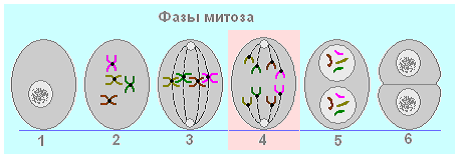
Mitosis– indirect division of a somatic eukaryotic cell, includes four phases. As a result of mitosis, the hereditary material of the mother cell is first doubled and then evenly distributed between the daughter cells. The duration of mitosis in animal cells is 30-60 minutes, and in plant cells - 2-3 hours.
Earthworm.
| Mitosis phase | Scheme | Amount of genetic material | Processes |
| Prophase | 2n 2chr 4c |
|
|
| Metaphase | 2n 2chr 4c |
|
|
| Anaphase | 2n 1chr 2c at each pole of the cell |
|
|
| Telophase | 2n 1chr 2c |
|
|
| 2n 1chr 2c |
|
15. As a result of meiosis,
- 2 daughter cells with a haploid set of chromosomes
- 2 daughter cells with a diploid set of chromosomes
- 4 daughter cells with a haploid set of chromosomes
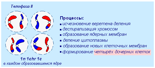
Telophase II. After completion of the first meiotic division, a short interphase of the second meiotic division follows. Moreover, at this stage, DNA replication (doubling) does not occur, and, therefore, diploidity is not restored.